Figures & data
Figure 1. Construction of DsbC-K2S fusion plasmid. (A) Identification of DsbC-K2S fusion plasmid by restriction digestion. Lane 1: 1-kb DNA marker. Lane 2: pET40b plasmid digested with BamHI and XhoI. Lane 3: DsbC-K2S plasmid digested with BamHI and XhoI. (B) Map of the DsbC- K2S fusion plasmid.
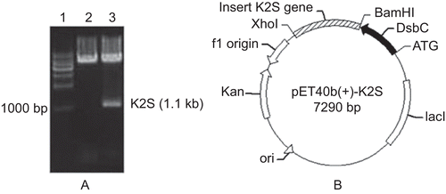
Figure 2. The optimal expression conditions on K2S expression. Lane 1: protein marker. Lane 2: the expression product of pET40b (+)-K2S induced by optimal expression conditions. Lane 3: the expression product of pET40b (+) vector.
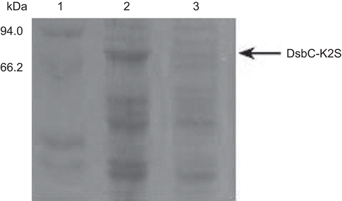
Figure 3. The elution curve of the Ni2+-chelating affinity chromatography. Fifty microliters of buffer NTA-0 was used to balance the column, then the sample was added. From 0 to 22.5 min was the penetration. After eluted with the elution buffer gradiently. The target protein was obtained at 46–73 min when the imidazole concentration reached at 10 mM.
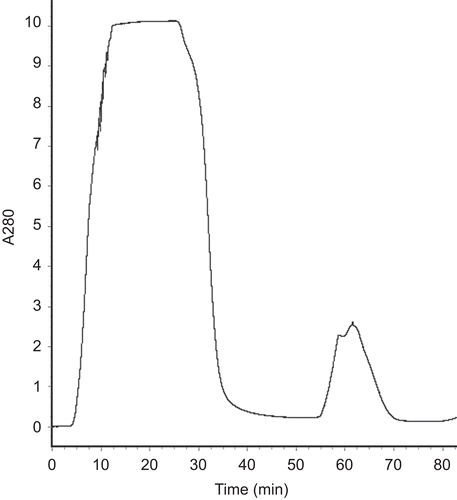
Figure 4. The SDS-PAGE of the purified DsbC-K2S fusion protein. Lane 1: protein marker; lane 2: the supernatant of the recombinant cell lysate; lane 3: penetration; lane 4: purified DsbC-K2S fusion protein.
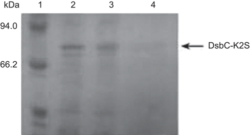
Figure 5. SDS-PAGE analysis of the cleaving of the fusion protein. Lane 1: DsbC-K2S fusion protein cleaved by Factor Xa. Lane 2: DsbC-K2S fusion protein. Lane 3: protein marker.
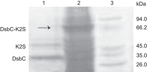
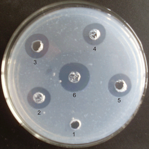
Figure 6. Thrombolytic activity assay of the recombinant K2S. 1: protein product of pET40b (+) vector. 2: the DsbC-K2S fusion protein (125 μg). 3 and 5: K2S protein after Factor Xa cutting (50 μg). 4: tPA (10 U). 6: Urokinase (10 U).