Figures & data
Figure 1. The chemical structure of ginsenoside Rb3.
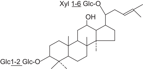
Figure 2. Effects of ginsenoside Rb3 on myocardial infarct size in myocardial ischemia-reperfusion injury in rats. The myocardial infarct size was stained by triphenyltetrazolium chloride. (A) Control group. (B) Myocardial ischemia-reperfusion group. (C) Ginsenoside Rb3 (5 mg/kg) group. (D) Ginsenoside Rb3 (10 mg/kg) group. (E) Ginsenoside Rb3 (20 mg/kg) group.
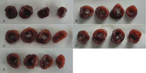
Figure 3. Effects of ginsenoside Rb3 on myocardial infarct size in myocardial ischemia-reperfusion injury in rats. Data were expressed as the mean ± SD (n = 8–10). Statistical significances were determined using one-way analysis of variance (ANOVA) followed by the least significant difference test. ##p < 0.01 compared with control group; *p < 0.05, **p < 0.01 compared with myocardial ischemia-reperfusion group.
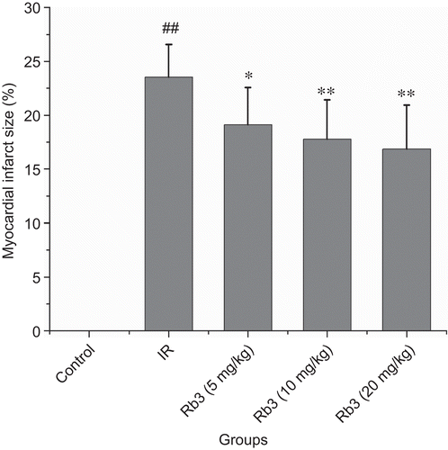
Figure 4. Effects of ginsenoside Rb3 on CK-MB activity in myocardial ischemia-reperfusion injury in rats. Data were expressed as the mean ± SD (n = 8–10). Statistical significances were determined using one-way analysis of variance (ANOVA) followed by the least significant difference test. ##p < 0.01 compared with control group; *p < 0.05, **p < 0.01 compared with myocardial ischemia-reperfusion group.
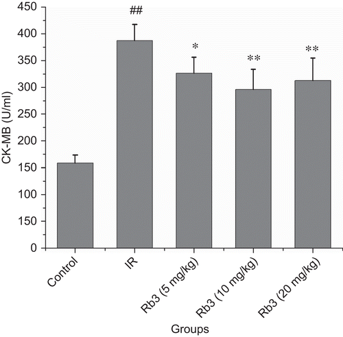
Figure 5. Effects of ginsenoside Rb3 on LDH activity in myocardial ischemia-reperfusion injury in rats. Data were expressed as the mean ± SD (n = 8–10). Statistical significances were determined using one-way analysis of variance (ANOVA) followed by the least significant difference test. ##p < 0.01 compared with control group; *p < 0.05, **p < 0.01 compared with myocardial ischemia-reperfusion group.
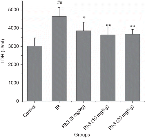
Figure 6. Effects of ginsenoside Rb3 on pathological changes of left ventricle in myocardial ischemia-reperfusion injury in rats. (H&E, 400×). (A) Control group showed clear integrity of myocardial cell membrane, and no inflammatory cell infiltration. (B) Myocardial ischemia-reperfusion group showed widespread myocardial structure disorder and subendocardial necrosis, which is related to infiltration with neutrophil granulocytes and myofibrillar fracture. (C) Ginsenoside Rb3 group showed mild necrosis and less infiltration of inflammatory cells. Bar = 400 μm.
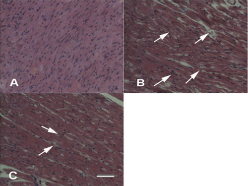
Figure 7. Effects of ginsenoside Rb3 on MDA content in myocardial ischemia-reperfusion injury in rats. Data were expressed as the mean ± SD (n = 8–10). Statistical significances were determined using one-way analysis of variance (ANOVA) followed by the least significant difference test. ##p < 0.01 compared with control group; *p < 0.05, **p < 0.01 compared with myocardial ischemia-reperfusion group.
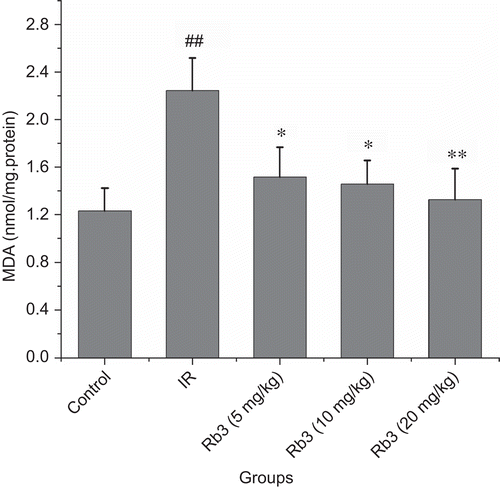
Figure 8. Effects of ginsenoside Rb3 on SOD activity in myocardial ischemia-reperfusion injury in rats. Data were expressed as the mean ± SD (n = 8–10). Statistical significances were determined using one-way analysis of variance (ANOVA) followed by the least significant difference test. ##p < 0.01 compared with control group; *p < 0.05, **p < 0.01 compared with myocardial ischemia-reperfusion group.
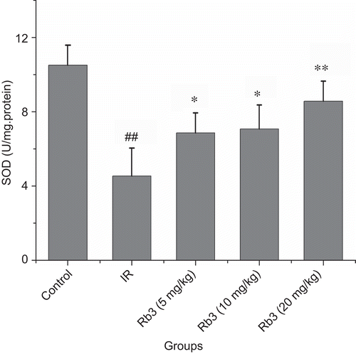
Figure 9. Effects of ginsenoside Rb3 on endothelin level in myocardial ischemia-reperfusion injury in rats. Data were expressed as the mean ± SD (n = 8–10). Statistical significances were determined using one-way analysis of variance (ANOVA) followed by the least significant difference test. ##p < 0.01 compared with control group; *p < 0.05, **p < 0.01 compared with myocardial ischemia-reperfusion group.
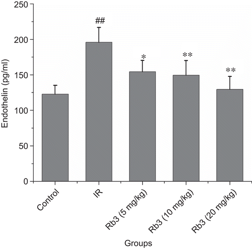
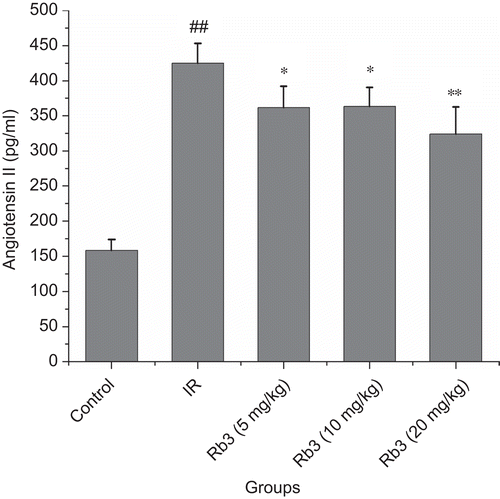
Figure 10. Effects of ginsenoside Rb3 on angiotensin II level in myocardial ischemia-reperfusion injury in rats. Data were expressed as the mean ± SD (n = 8–10). Statistical significances were determined using one-way analysis of variance (ANOVA) followed by the least significant difference test. ##p < 0.01 compared with control group; *p < 0.05, **p < 0.01 compared with myocardial ischemia-reperfusion group.