Figures & data
Figure 1. A synthetic scheme map of salvianic borneol ester (SBE).
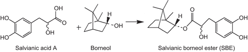
Figure 2. The destabilization of Aβ1–40 oligomers measured by the thioflavin T fluorescence assay. Preformed Aβ1–40 oligomers were incubated in the presence of salvianic borneol ester (SBE) (10 µg/mL), borneol (10 µg/mL), salvianic acid A (10 µg/mL), or the mixture of borneol and salvianic acid A (1:1, both 5 µg/mL) for 30 min. Values are reported as mean ± SD (n = 3). *P < 0.05, **P < 0.001 versus control (Aβ1–40 oligomers without treatment) (one-way ANOVA analysis with Dunnett’s post hoc test).
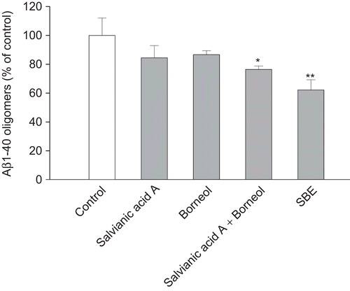
Figure 3. Effects of salvianic borneol ester (SBE) on Aβ1–40 oligomers destabilization measured by the thioflavin T fluorescence assay. SBE destabilized Aβ1–40 oligomers (A) in dose-dependent manner (incubation for 30 min) and in time-dependent manner (SBE at 10 µg/mL) (B). Values are reported as mean ± SD (n = 3). Significant destabilizing activities of SBE were observed dose-dependently at concentration above 5 µg/mL (P < 0.001 vs. control) and time-dependently at and above 1 h (P < 0.001 vs. control) (one-way ANOVA analysis with Dunnett’s post hoc test).
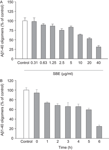
Figure 4. Electron micrographs of Aβ1–40 oligomers. Aβ1–40 oligomers were incubated in the absence (A) or presence of salvianic borneol ester (SBE) 2.5 µg/mL (B), 20 µg/mL (C), or 40 µg/mL (D). Magnification: 100,000×; scale bar = 170 nm.
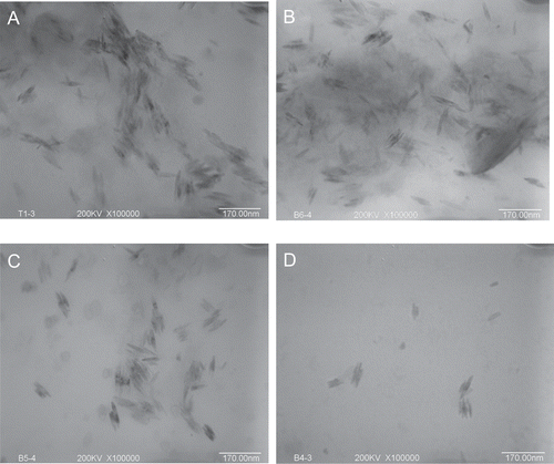
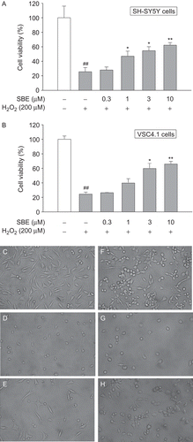
Figure 5. The protection of salvianic borneol ester (SBE) against toxicity induced by H2O2 in SH-SY5Y cells and VSC 4.1 cells. MTT cell viability assay for SH-SY5Y cells (A) and VSC 4.1 cells (B). Values are reported as mean ± SD (n = 4–6). *P < 0.05, **P < 0.001 versus H2O2 alone. ##P < 0.001 versus control (one-way ANOVA analysis with Dunnett’s post hoc test). Representative photomicrographs showing SH-SY5Y cells exposed to vehicle (C), H2O2 (D), or both H2O2 and 10 µM SBE (E); VSC 4.1 cells exposed to vehicle (F), H2O2 (G), or both H2O2 and 10 µM SBE (H). Magnification: 400×.