1,299
Views
20
CrossRef citations to date
0
Altmetric
Research Article

Silymarin selectively protects human renal cells from cisplatin-induced cell death

, , &
Pages 1082-1090 | Received 19 Nov 2010, Accepted 01 Mar 2011, Published online: 18 May 2011

Figures & data

Figure 1.  Cytotoxic effect of cisplatin on HK-2, H460, and G361 cells. (A–C) Cells were treated with various concentrations of cisplatin (0–300 µM) for 24 h and cell viability was measured by MTT assay. Values are means ± SD of three independent experiments, *P < 0.05 versus non-treated control. (D–F) Apoptotic and necrotic cells were detected by Hoechst 33342 and propidium iodide co-staining assay and examined under a fluorescence microscope.
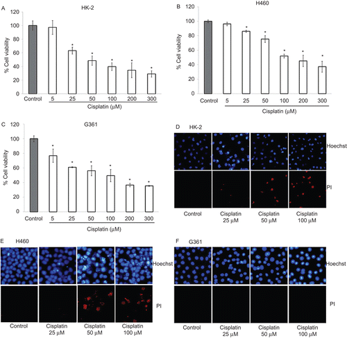
Figure 1.  Cytotoxic effect of cisplatin on HK-2, H460, and G361 cells. (A–C) Cells were treated with various concentrations of cisplatin (0–300 µM) for 24 h and cell viability was measured by MTT assay. Values are means ± SD of three independent experiments, *P < 0.05 versus non-treated control. (D–F) Apoptotic and necrotic cells were detected by Hoechst 33342 and propidium iodide co-staining assay and examined under a fluorescence microscope.

Figure 2.  Effect of silymarin on HK-2, H460, and G361 cells. (A–C) Cells were treated with various concentrations of silymarin (0–200 µM) for 24 h and cell viability was measured by MTT assay. Values are means ± SD of three independent experiments, *P < 0.05 versus non-treated control. (E–F) Nuclear morphology of apoptosis and necrosis detected by Hoechst 33342 and propidium iodide assay.
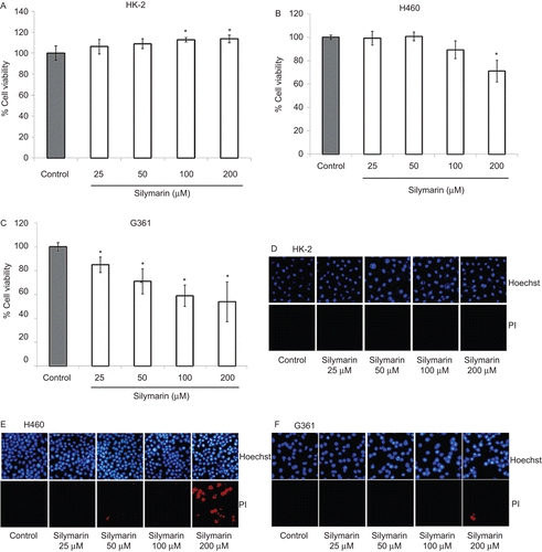
Figure 2.  Effect of silymarin on HK-2, H460, and G361 cells. (A–C) Cells were treated with various concentrations of silymarin (0–200 µM) for 24 h and cell viability was measured by MTT assay. Values are means ± SD of three independent experiments, *P < 0.05 versus non-treated control. (E–F) Nuclear morphology of apoptosis and necrosis detected by Hoechst 33342 and propidium iodide assay.
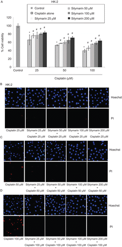
Figure 3.  Protective effect of silymarin on cisplatin-induced HK-2 cell death. (A) Cells were pretreated with various concentrations of silymarin (25–200 µM) for 1 h prior to 24-h cisplatin exposure. Cell viability was measured by MTT assay. Values are means ± SD of three independent experiments, *P < 0.05 versus non-treated control, and #P < 0.05 versus cisplatin-treated control. (B–D) Nuclear morphology of apoptosis and necrosis detected by Hoechst 33342 and propidium iodide assay.
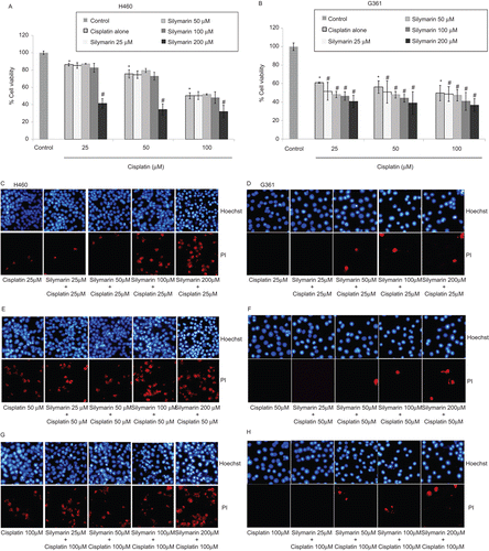
Figure 3.  Protective effect of silymarin on cisplatin-induced HK-2 cell death. (A) Cells were pretreated with various concentrations of silymarin (25–200 µM) for 1 h prior to 24-h cisplatin exposure. Cell viability was measured by MTT assay. Values are means ± SD of three independent experiments, *P < 0.05 versus non-treated control, and #P < 0.05 versus cisplatin-treated control. (B–D) Nuclear morphology of apoptosis and necrosis detected by Hoechst 33342 and propidium iodide assay.

Figure 4.  Effect of silymarin on cisplatin-induced H460 and G361 cell death. (A and B) Cells were pretreated with various concentrations of silymarin (25–200 µM) for 1 h prior to 24-h cisplatin treatment. Cell viability was measured by the MTT assay. Values are means ± SD of three independent experiments, *P < 0.05 versus non-treated control, and #P < 0.05 versus cisplatin-treated control. (C–H) Nuclear morphology of apoptosis and necrosis detected by Hoechst 33342 and propidium iodide assay.

Figure 4.  Effect of silymarin on cisplatin-induced H460 and G361 cell death. (A and B) Cells were pretreated with various concentrations of silymarin (25–200 µM) for 1 h prior to 24-h cisplatin treatment. Cell viability was measured by the MTT assay. Values are means ± SD of three independent experiments, *P < 0.05 versus non-treated control, and #P < 0.05 versus cisplatin-treated control. (C–H) Nuclear morphology of apoptosis and necrosis detected by Hoechst 33342 and propidium iodide assay.

Reprints and Corporate Permissions

Please note: Selecting permissions does not provide access to the full text of the article, please see our help page How do I view content?

To request a reprint or corporate permissions for this article, please click on the relevant link below:

Academic Permissions

Please note: Selecting permissions does not provide access to the full text of the article, please see our help page How do I view content?

Obtain permissions instantly via Rightslink by clicking on the button below:

If you are unable to obtain permissions via Rightslink, please complete and submit this Permissions form. For more information, please visit our Permissions help page.