Figures & data
Figure 1. Histological sections of cutaneous wound site obtained from the controls, HOT and HOTBp lesions of the rats (A) normal histological skin tissue in the first day. (H&E, ×200) (B) normal histological skin tissue in the first day samples stained with Gomori trichrome stain (×200); (C) Late phase granulation tissues which were characterized with fibroblastic proliferation and scattered lymphocytes in a fifth day subject. Normal dermal tissue is seen at right corner [(rectangular) (Hematoxylin and eosin, ×100); (D) The collagen fibers started to emerge slightly in the granulation tissue in the 5th day samples (Gomori trichrome stain, ×100); (E) a lesion at a 10th day subject. Collagen fibrils is seen as more abundant than the 10th day sample (Gomori trichrome stain, ×100), (F) a sample of control lesion in the 30th day. Healing with fibrous scar formations is seen (Hematoxylin and eosin, ×100) (G) same tissue of F with Gomori trichrome stain (×100) (H, J) Near normal skin tissue without fibros scar tissue seen in a 30th day sample treated with Bellis perennis (Hematoxylin and eosin, Gomori trichrome stain, respectively, ×100). (e) epidermis, (d) dermis, (ndt) normal dermal tissue, (cf) collagen fibers.
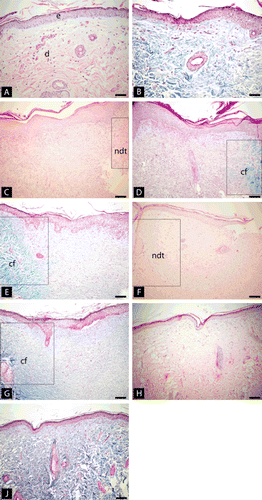
Figure 2. Wound closure percentages of the control, HOT and HOTBp groups at the 5th, 10th and the 30th days of the experiment.
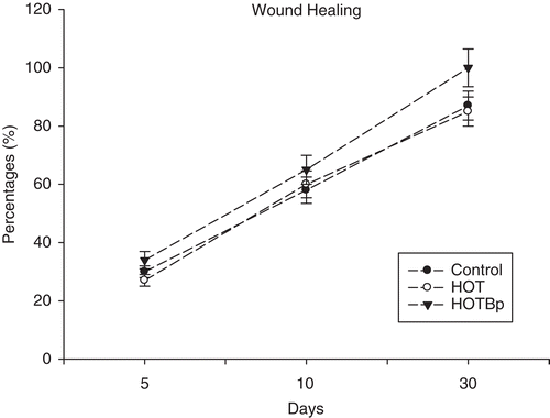
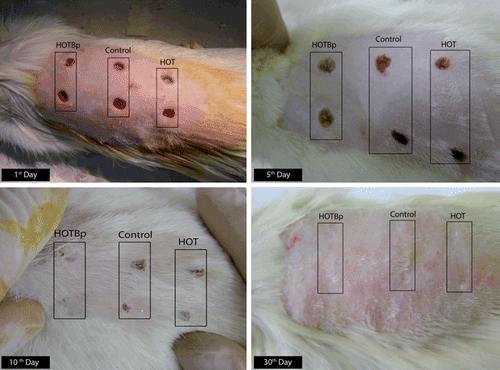
Figure 3. Photographs of the different rats in different days of the experiment. HOT: hydrophilic ointment treatment, HOTBp: hydrophilic ointment loaded with B. perennis fraction.