Figures & data
Figure 1. Chemical structure of 4-acetoxy-plakinamine B.
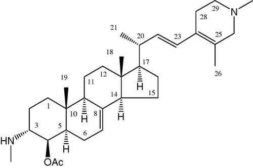
Figure 2. Electrostatic surface of AChE showing the aromatic gorge (active site) of enzyme: (A) lateral view of AChE. Encircled regions depict the aromatic gorge; (B) side view (at 90°) of AChE; (C) a closer side view of the aromatic gorge of AChE along with bound inhibitor (4APB) revealing molecular interactions between inhibitor and amino acid residues of surrounding active site of AChE. Surface colors red (aggregated negatively charged area), blue (aggregated positively charged area) and white (neutral or hydrophobic area) colors are shown.
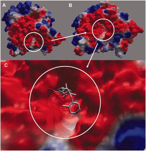
Figure 3. Molecular interactions of 4APB with selected amino acid residues inside the active site of AChE. Hydrogen atoms are omitted (except polar ones) for the purpose of clarity.
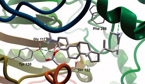
Figure 4. A closer view of the binding mode of 4APB inside the active site of AChE. Hydrogen atoms are omitted (except polar ones) for clarity. Hydrogen bonding is shown in dotted lines.
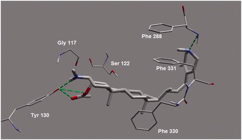
Figure 5. A 3D-cut view showing electrostatic surface of the binding pocket. ES and AP subsites are shown as a deeper pocket while PAS is shown toward the viewer.
