Figures & data
Figure 1. Effect of Saussurea lappa extract on cell viability in KB cells. Cells were treated with various concentrations of Saussurea lappa extract for 24 h in KB cells (A). 30 µg/ml of Saussurea lappa extract treated into cells for different time periods (B). Cell viabilities were determined by the MTT assay. The percentage of cell viability was calculated as a ratio of A570 nm. Results were expressed as percent of the control. Each data point represents the mean ± SEM from three experiments.
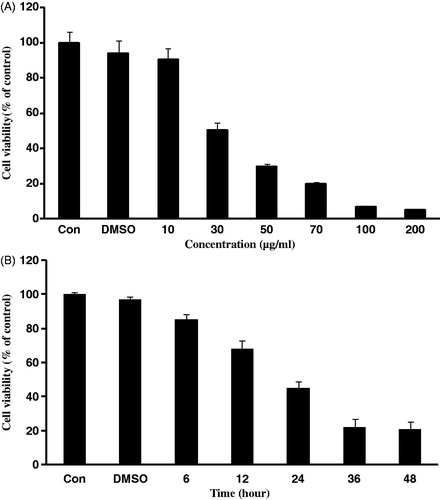
Figure 2. The expression levels of apoptosis-related proteins by treatment with Saussurea lappa extract in KB cells. KB cells were seeded at 1 × 10⁶ and were then treated with 30 µg/ml of Saussurea lappa extract for the indicated time point (12 h and 24 h). Fragmentation of internucleosomal DNA by Saussurea lappa extract treatment in KB cells. Genomic DNA was subjected to 1.5% agarose gel electrophoresis (A). After 12 h, and 24 h Saussurea lappa extract treatment, mRNA was determined by RT-PCR (B), and Bax and Bcl-2 protein levels were determined by Western blot analysis (C). Whole cell lysates were separated by 12% SDS-PAGE and probed for Bax, Bcl-2 and β-actin as a loading control.
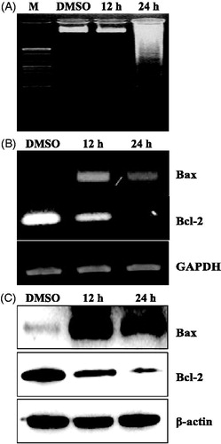
Figure 3. Saussurea lappa extract caused caspase-3-dependent apoptosis. Caspase-9, cleaved caspase-9, caspase-3, cleaved caspase-3, PARP and cleaved PARP protein levels were determined by western blot analysis. Whole-cell lysates (50 µg/lane) were subjected to immunoblotting for the indicated proteins. Probing with β-actin was used to show equal protein loading (A). Activation of caspase-3/7 by Saussurea lappa extract treatment in living KB cells. The cells were treated with 30 μg/ml of Saussurea lappa extract for 24 h and followed by adding specific cell-permeable substrate Phiphilux G1D2. Caspase-3/7 activity was visualized by fluorescence microscopy (B).
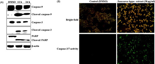
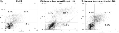
Figure 4. Saussurea lappa extract induced apoptosis in KB cells. Cells were treated with 30 µg/ml of Saussurea lappa extract for 12 h and 24 h. The cells were stained with Annexin V-FITC and propidium iodine (PI). The apoptotic cells were then analyzed by fluorescence-activated cell sorting (FACS) analysis. This apoptotic data was determined by FACS analysis showing the percentages of lower right quadrant for early and upper right quadrant for late apoptotic cells.