Figures & data
Figure 1. Act1 mRNA expressed in synovioblast cells and SW982 cells. (A) Gel image of RT-PCR product for Act1 mRNA. Lines 1, 2: Act1 mRNA (415 bp) in primary cultured RA fibroblast-like synovial cells; line 3: Act1 mRNA in SW982 cells; lines 4–6: β-actin mRNA (220 bp) in SW982 cells; M: Trans DNA Marker I. (B) Representative Western blot bands of Act1 and β-actin are shown. Line 1: SW982; lines 2–3: primary cultured RA fibroblast-like synovial cells.
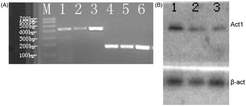
Figure 2. Act1 mRNA levels are induced by different concentrations and different incubation times of IL-17 in SW982 cells. (A) Gel image of RT-PCR product for Act1 mRNA 4 h after stimulation by IL-17. Lines 1–6: Act1 mRNA in 100, 50, 20, 10, 5, 0 ng/ml IL-17; lines 1′–6′: the corresponding β-actin mRNA. (B) The quantitive analysis for Act1 mRNA in SW982 cells. The Act1 mRNA level was increased with the elevated concentration of IL-17. The data are expressed as mean ± SEM (n = 9 per group). **p < 0.01, ***p < 0.001, compared with 0 ng/ml IL-17. (C) Gel image of RT-PCR product for Act1 mRNA after stimulation by 20 ng/ml IL-17. Lines 1–6: Act1 mRNA in 0 min, 15 min, 30 min, 1 h, 2 h, 4 h after induction by IL-17; lines 1′–6′-: the corresponding β-actin mRNA. (D) The quantitative analysis for Act1 mRNA in SW982 cells. The Act1 mRNA level was increased with the stimulation time of IL-17. The data are expressed as mean ± SEM (n = 9 per group). *p < 0.05, **p < 0.01, ***p < 0.001, compared with IL-17 at 0 min.
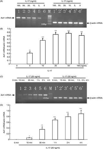
Figure 3. Traf3ip2 siRNA significantly decreased Act1 protein expression level in SW982 cells. (A) Representative Western blot bands of Act1 are shown. Lines 1–2: control-siRNA; line 3: Traf3ip2-siRNA. (B) The DY-547-labeled SW982 cells after transfection with Traf3ip2 gene for 48 h. Left: common microscopy; right: fluorescence microscopy (×400).
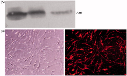
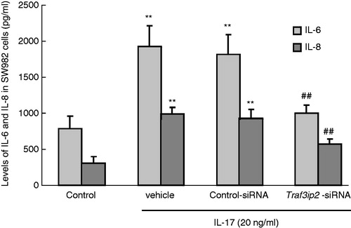
Figure 4. IL-6 and IL-8 were induced in SW982 cells with various treatments. IL-17 (20 ng/ml) significantly increased IL-6 and IL-8 in SW982 cells, while transfection of Traf3ip2 siRNA markedly decreased IL-6 and IL-8 level compared to transfection control-siRNA group. The data are expressed as mean ± SEM (n = 9 per group). **p < 0.01 compared with the control group; ##p < 0.01 compared with the control-siRNA group.