Figures & data
Figure 1. Liver glycogen reserves in experimental animals during NDMA-induced hepatic fibrosis and subsequent treatment with vitamin B12. Values are expressed as mean ± SD (*p < 0.05, **p < 0.001).
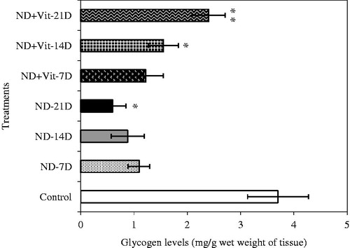
Table 1. Changes in biochemical parameters of hepatic injury in the sera and liver of control and treated rats.
Figure 2. Vitamin B12 levels in the liver and plasma of experimental animals during NDMA-induced hepatic fibrosis and after treatment with vitamin B12. Values are expressed as mean ± SD (*p < 0.05, **p < 0.001).
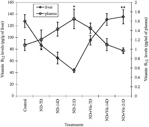
Figure 3. Hematoxylin and eosin (H & E) staining of rat liver during NDMA-induced hepatic fibrosis and subsequent treatment with vitamin B12. (A) Control liver showing central vein and characteristic lobular architecture (10 × ). (B) Day 7. Peripheral lymphocyte infiltration (4×). (C) Day 14. Demonstrating Kupffer cell hyperplasia (40×). (D) Day 21. Liver congestion, lymphocyte infiltration and well developed fibrosis (10×). (E) Control liver with normal structure subsequent to vitamin B12 treatment only (4×). (F) Day 7. Liver showing lymphocyte and neutrophilic infiltration with mild fibrosis (40×). (G) Day 14. Inflammation absent with decreased fibrosis (10×). (H) Day 21. Restoration of normal liver architecture with increasing density of regenerating hepatocytes (10×).
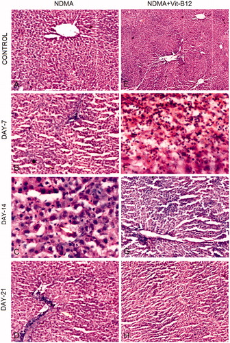
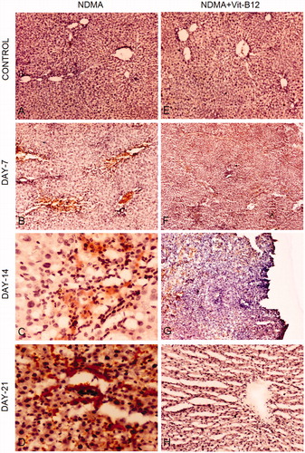
Figure 4. Immunohistochemical staining for α-SMA during the pathogenesis of NDMA-induced hepatic fibrosis and subsequent treatment with vitamin B12. (A) Normal liver with central vein (4×). (B) Day 7. Focal α-SMA staining around central vein positively stained stellate cells (10×). (C) Day 14. Focal positivity and α-SMA stained regions showing widespread activation of hepatic stellate cells (40×). (D) Day 21. Stellate cells showing intense focal staining of α-SMA in fibrotic region (40×). (E) Normal liver architecture in vitamin B12 treated rats (4×). (F) Day 7. Focal positivity of α-SMA (4×). (G) Day 14. Scattered and decreased focal positive staining of α-SMA showing reduced number of activated hepatic stellate cells (10×). (H) Day 21. Regenerating hepatocytes with scant positivity around central vein (4×).