
Figures & data
Table 1. Chemical composition of the essential oil of flowers of M. suaveolens Ehrh. cultivated in Egypt.
Figure 1. Photographs of Mentha suaveolens Ehrh. (A) A flowering branch of Mentha suaveolens Ehrh. (X = 0.6). (B) The inflorescence (X = 1). (C) The bract (X = 10). (D) The flower (X = 12).
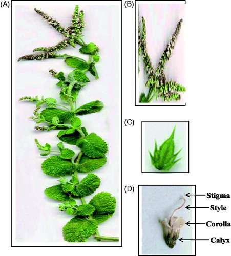
Figure 2. Transverse section of the rachis of the inflorescence of Mentha suaveolens Ehrh. (A) Low power view (X = 30). (B) High power view (X = 215). (C) High power view from the corners (X = 315). col., collenchyma; c.par., cortical parenchyma; end., endodermis; ep., epidermis; m.r., medullary ray; n.gl.t., non-glandular trichomes; per., pericycle; ph., phloem; pi., pith; xy., xylem.
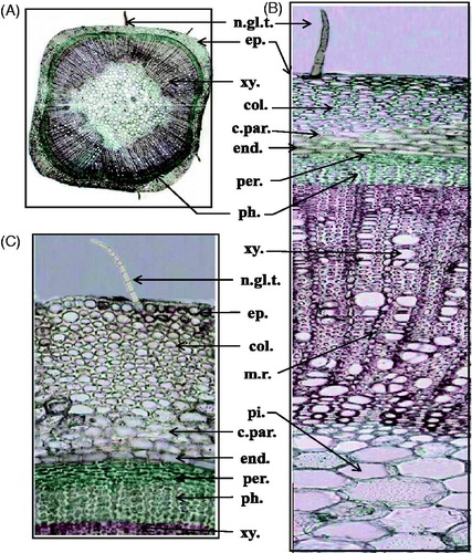
Figure 3. Transverse section of the bract of Mentha suaveolens Ehrh. (A) Low power view (X = 40). (B) High power view of the midrib (X = 100). (C) High power view of the lamina (X = 333). c.par., cortical parenchyma; ca., cambium; gl.t., glandular trichomes; l.ep., lower epidermis; m.r., medullary ray; n.gl.t., non-glandular trichomes; pal.t., palisade tissue; per., pericycle; ph., phloem; sp.t., spongy tissue; u.ep., upper epidermis; xy., xylem.
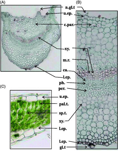
Figure 4. Epidermal cells of the rachis and bract of Mentha suaveolens Ehrh. (A) Epidermal cells of the rachis (X = 450). (B) Upper epidermal cells of the bract (X = 433). (C) Lower epidermal cells of the bract (X = 433). cap.gl.t., capitate glandular trichomes; cic., cicatrix; lab.gl.t., labiaceous glandular trichomes; str.cu., striated cuticle.
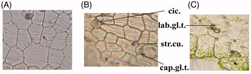
Figure 5. Transverse section of the flower of Mentha suaveolens Ehrh. (At the upper part of the calyx) (X = 60). ant., anther; pet., petals; p.gr., pollen grains; sep., sepals; sty., style.
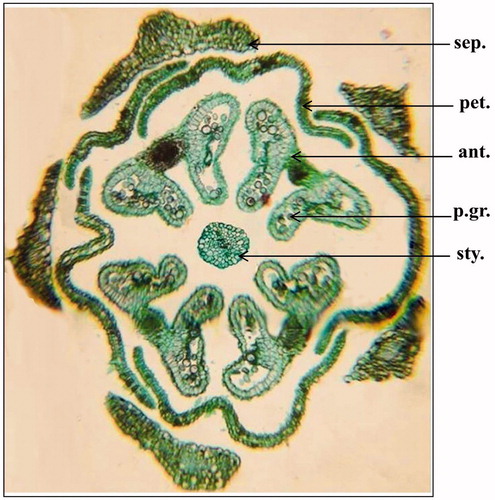
Figure 6. Micromorphology of the calyx and corolla of Mentha suaveolens Ehrh. (A) Transverse section of the sepal (X = 60). (B) Inner epidermal cells of the calyx at the base (X = 450). (C) Inner epidermal cells of the calyx at the tip (X = 450). (D) Outer epidermal cells of the calyx at the base (X = 450). (E) Outer epidermal cells of the calyx at the tip (X = 450). (F) Transverse section of the petal (X = 140). (G) Inner epidermal cells of the corolla at the base (X = 775). (H) Inner epidermal cells of the corolla at the tip (X = 775). (I) Outer epidermal cells of the corolla at the base (X = 775). (J) Outer epidermal cells of the corolla at the tip (X = 775). c.par., cortical parenchyma; l.ep., lower epidermis; n.gl.t., non-glandular trichomes; u.ep., upper epidermis; v.b., vascular bundle.
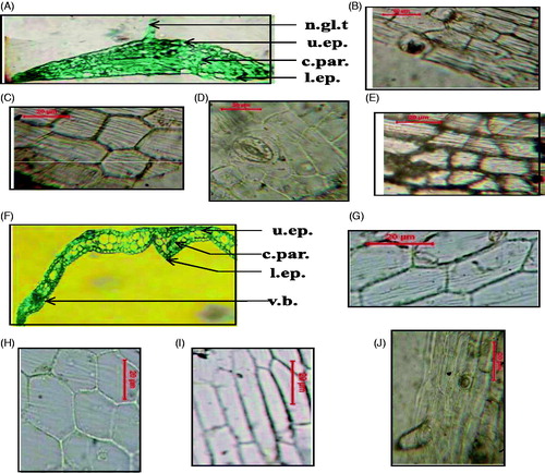
Figure 7. Micromorphology of the androecium and gynaecium of Mentha suaveolens Ehrh. (A) Detailed transverse section of the anther (X = 160). (B) Detailed transverse section of the filament (X = 125). (C) Detailed transverse section of the ovary (X = 160). (D) Ovary wall (X = 333). (E) Transverse section of the style (X = 267). (F) Transverse section of the stigma (X = 217). c.par., cortical parenchyma; c.t., connective tissue; ep., epidermis; f.l., fibrous layer of anther; i.ep., inner epidermis; n.gl.t., non-glandular trichome; o.ep., outer epidermis; ov., ovule; o.w., ovary wall; p.gr., pollen grains; p.s., pollen sac; v.b., vascular bundle.
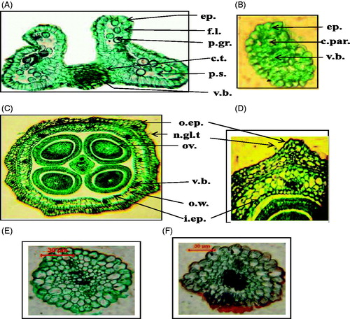
Figure 8. Powdered inflorescence of Mentha suaveolens Ehrh. ep.an, epidermis of anther (X = 650); ep.br, epidermis of bract (X = 433); ep.ca, epidermis of calyx (X = 450); ep.cr, epidermis of corolla (X = 775); ep.fil, epidermis of filament (X = 650); ep.ov., epidermal cells of the ovary (X = 375); ep.ra, epidermis of rachis (X = 450); ep.stg., epidermal cells of the stigma (X = 450); ep.sty., epidermal cells of the style (X = 525); f.l., fibrous layer of anther (X = 650); gl.t., glandular trichomes (X = 433); m.r., medullary ray (X = 550); n.gl.t., non-glandular trichomes (X = 433); p.gr., pollen grains (X = 650); pal., palisade cells (X = 400); v.o.dp., volatile oil droplets (X = 300); w.f., wood fibers (X = 233); w.p., wood parenchyma (X = 500); x.v., xylem vessel (X = 750).
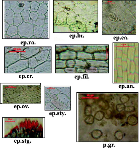
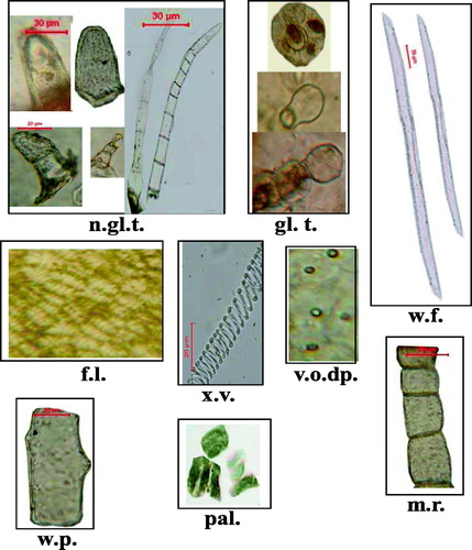
Table 2. Microscopical measurements of the different organs of the inflorescence M. suaveolens Ehrh.