
Figures & data
Figure 1. Scavenging effect of various concentrations of Origanum vulgare and BHT on the DPPH free radical, measured at 517 nm.
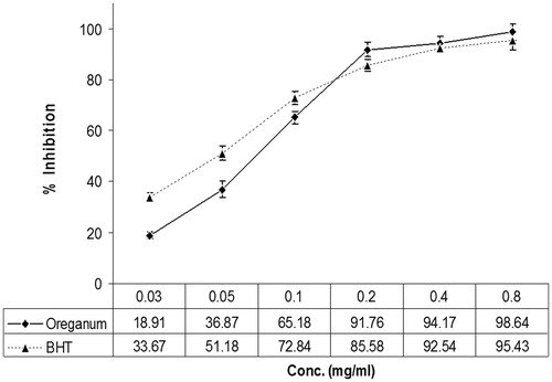
Table 1. Effects of CP (200 mg/kg) and/or OV on lung lipid peroxide formation expressed as TBARS, GSH content, and SOD activity.
Figure 2. Normal group: a section of the mouse lung tissue showing alveolar septa lined with normal pneumocytes (A, yellow arrow) and normal alveolar spaces (B, red arrow). (Hematoxylin and eosin-stained paraffin sections; H&E × 400.)
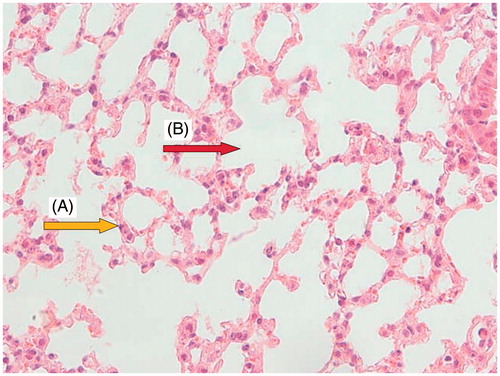
Figure 3. CP (200 mg/kg) group: a section of mouse lung showing hyperplastic pneumocytes (A, yellow arrow), respiratory type epithelium (B, green arrow), large distal air spaces filled by lymphocytes, neutrophils, and cell debris (C, red arrow). (Hematoxylin and eosin-stained paraffin sections; H&E × 400.)
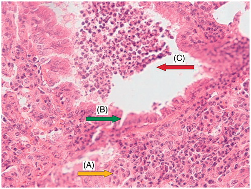
Figure 4. Group pretreated with 400 mg/kg Origanum for 7 d before CP administration: a section of mouse lung tissue showing remnants of neutrophilic infiltration and cell debris (A, green arrow), normal alveolar space (B, red arrow), and alveolar septa showing focally hyperplastic pneumocytes and RBC extravasation (C, yellow arrow). (Hematoxylin and eosin-stained paraffin sections; H&E × 400.)
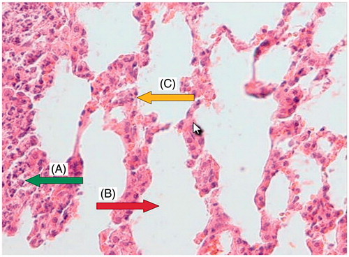
Table 2. The results of semi-quantitative histopathological examination of the lung, showing protection against CP-induced tissue damage by OV at various doses.