Figures & data
Figure 1. Effect of AMP on the viability of macrophages. The cells were treated with or without AMP (1–200 μg/mL) or 100 ng/mL LPS for 24 h. Cells were then assessed for viability using MTT.
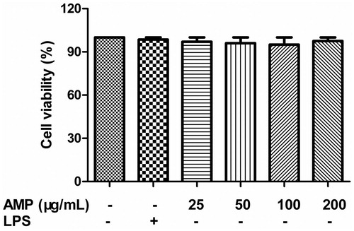
Figure 2. Effect of AMP on phagocytosis of macrophages. Macrophages (1 × 10⁶ cells/mL) were preincubated with different concentrations of AMP (25, 50, 100, and 200 μg/mL) or 100 ng/mL LPS, and then incubated with FITC-dextran (1 mg/mL) at 37 °C for 1 h. The extent of the phagocytic uptake was determined by flow cytometry. *p < 0.05.
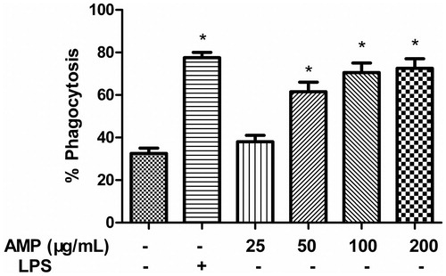
Figure 3. NO, TNF-α and IFN-γ production in macrophages incubated with AMP. Macrophages were incubated with different concentrations of AMP (25, 50, 100, and 200 μg/mL) or 100 ng/mL LPS for 24 h. Supernatants were harvested and the level of NO, TNF-α, and IFN-γ was determined by the Griess reagent and ELISA. *p < 0.05.
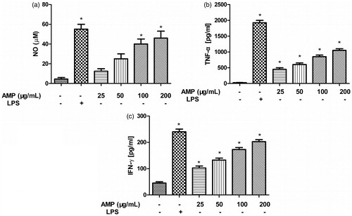
Figure 4. IκB degradation and activation of NF-κB in macrophages treated with AMP. Macrophages were stimulated with different concentrations of AMP for 4 h. (a and c) IκB degradation and activation of NF-κB was analyzed by Western blotting with anti-IκB and anti-NF-κB p65 antibodies. (b and d) Expression of IκB and NF-κB p65 was quantified. The ratios for these proteins are shown. *p < 0.05 compared with the untreated group.
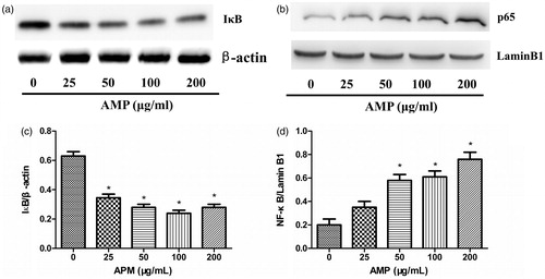
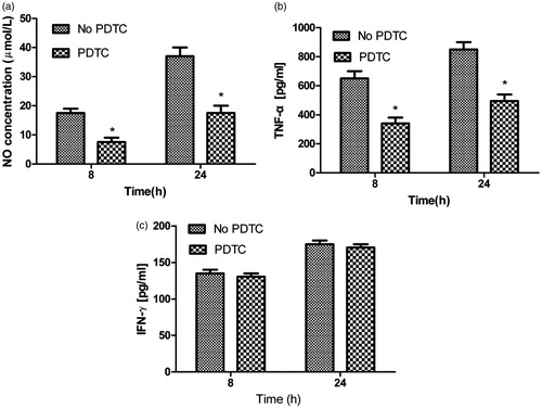
Figure 5. Effect of NF-κB inhibitor on NO, TNF-α, and IFN-γ production in macrophages incubated with AMP. Macrophages were pretreated with PDTC (100 μM) for 90 min, and then stimulated by AMP. Supernatants were harvested and the level of NO (a), TNF-α (b), and IFN-γ (c) was determined by ELISA and Griess reagent.