Figures & data
Figure 1. Effects of ID1216 on the adipose tissue and liver. Mice fed an HF diet for 10 weeks were sacrificed, and epididymal adipose tissue and liver weights were measured (A). Data are expressed as means ± SEM of 8 mice per group. *p < 0.05, **p < 0.01 versus HF. Sections of epididymal adipose tissues and livers were subjected to hematoxylin and eosin (H&E) staining and then analyzed by microscopy (B). H&E were stained sections of epididymal adipose tissues and liver from each group showing the various degrees of lipid droplets formation.
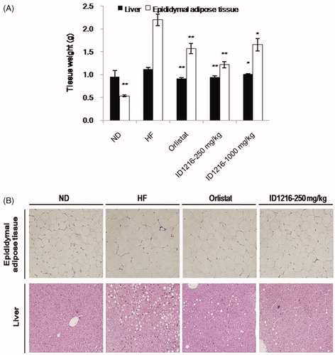
Table 1. Effects of ID1216 on body weight, food intake, fecal triglyceride, and plasma lipids in mice fed a HF diet.
Figure 2. Effects of ID1216 on adaptive thermogenesis and glucose tolerance in diet-induced obese mice. Mice fed an HF diet for 7 weeks were exposed to 4 °C and rectal temperature was monitored for 8 h (A). Data are expressed as means ± SEM of 6 mice per group. *p < 0.05, **p < 0.01 versus HF. Blood glucose concentrations (B) and AUC (C) during the OGTT and the fasting plasma glucose (D). Data are expressed as mean ± SEM of 8 mice per group. *p < 0.05, **p < 0.01 versus HF.
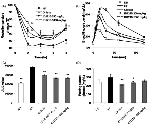
Figure 3. Inhibition of adipocyte differentiation by ID1216. 3T3-L1 cells were differentiated for 14 d with or without ID1216 (50 μg/ml). Oil red O-stained dishes and high magnification (× 200) of cells (A) and triglyceride normalized to total cellular protein (B) were observed the content of lipid accumulation from each group. Data are expressed as means ± SEM (n = 3). *p < 0.05 versus the vehicle group.
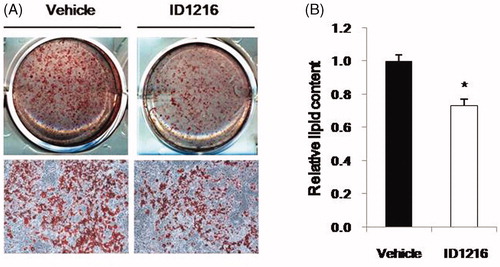
Table 2. Effect of ID1216 on the expression of key transcription factors during 3T3-L1 adipocyte differentiation.
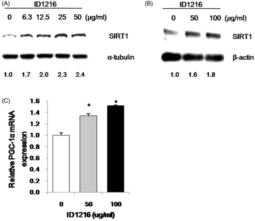
Figure 4. Effects of ID1216 on the expression of SIRT1 and PGC-1α. HEK293 cells (A) or C2C12 myocytes (B) were treated with various concentrations of ID1216 and extracted proteins after the mentioned time, respectively. The protein expression level of SIRT1 was detected by Western blot analysis and the relative ratio is shown. C2C12 myocytes were treated with or without ID1216 for 30 h and relative mRNA level was analyzed by quantitative RT-PCR (C). The values indicated relative mRNA levels compared with the vehicle group. Data are means ± SEM (n = 3). *p < 0.05, **p < 0.01 versus the non-treated group.