Figures & data
Table 1. Combination effect of Moringa oleifera along with piperine and curcumin against beryllium induced oxidative stress.
Table 2. Combination effect of Moringa oleifera along with piperine and curcumin against beryllium induced oxidative stress and disturbance in carbohydrate metabolism.
Figure 1. Photomicrograph of liver after subchronic exposure to beryllium followed by combination therapy of moringa oleifera. (A) Control rats showing well-formed chord arrangement of hepatocytes and central vein (HE, 400×). (B and C) Beryllium-treated rats showing central camal with debris, loss of chord arrangement, nuclei of hepatocytes are irregular, and hyperchromatic (HE, 100×, 400×). (D) Therapy with MO 150 mg showing large vesicular nuclei and proliferation of camamiculi (HE, 400×). (E) Therapy with MO+Pip showing congested central camal (HE, 400×). (F) Therapy with MO+Cur showing histoarchitecture of liver (HE, 400×).
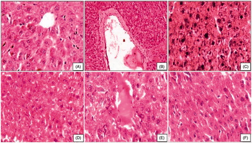
Figure 2. Photomicrograph of kidney after subchronic exposure to beryllium followed by combination therapy of moringa oleifera. (A) Control rats showing well-formed uriniferous tubules (HE, 400×). (B and C) Beryllium-treated rats showing obliteration of lumen of uriniferous tubules, exfoliation of nuclei, and contraction of glomerulus (HE, 400×). (D) Therapy with MO 150 mg showing uriniferous tubules with lumen and epithelial cell are vacuolated (HE, 400×). (E) Therapy with MO+Pip showing well-formed glomerulus and better lumen of uriniferous tubules (HE, 400×). (F) Therapy with MO+Cur showing well-formed glomeruli (HE, 400×).
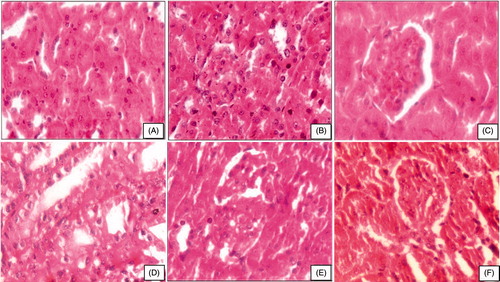
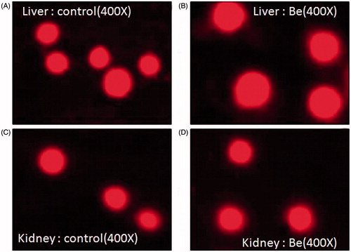
Figure 3. Evaluation of DNA damage by comet assay.