Figures & data
Figure 1. Chemical structure of diterpenoids 1–5 from seeds of Vietnamese C. sappan.
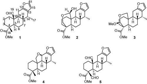
Table 1. Cytotoxic activity of compounds 1–5 from the seed of C. sappan.
Figure 2. The increment of caspase-3 activity by compound 3 in HL-60 cells. After 12 h, 24 h, and 48 h incubation with the 3-treated HL-60 cells, the cell lysates were incubated at 37 °C with caspase-3 substrate (Ac-DEVD-AFC) for 1 h. The fluorescence intensity of the cell lysates was measured to determine the caspase-3 activity. The blank group was used as 0.1% DMSO-treated cells; camptothecin (7.8 µM) was used as a positive control. Data are presented as the mean ± SD of results from three independent experiments.
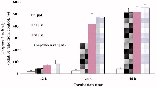
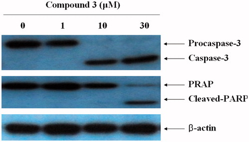
Figure 3. The induction of caspase-3 activation and PARP degradation by compound 3 in HL-60 cells. Western blot analysis of caspase-3 and PARP protein levels after exposure to compound 3. HL-60 cells were treated with compound 3 at the indicated concentrations. Protein (50 μg) from each sample was resolved by SDS-PAGE (10% (w/v) polyacrylamide gel).