Figures & data
Figure 1. DPPH free radical scavenging capacity of WEHDW and vitamin C.
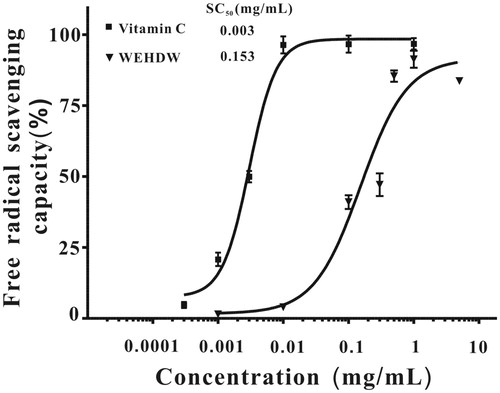
Figure 2. Examination of the cytotoxicity of H2O2 and the protective effect of WEHDW against H2O2-induced damage on LO2 cells. (A) Cells were treated with different concentrations (50, 100, 150, 200, 250, 300, 400, or 500 μM) of H2O2 for 6 h. Data were expressed as mean ± SEM of three separate experiments; ###p < 0.001 versus control. (B) LO2 cells were pretreated with WEHDW (0.3, 0.5, 1, 3, 5, or 10 mg/mL, 2 h) before challenging with H2O2 (200 μM, 6 h). Data were expressed as the means ± SEM of three separate experiments; ###p < 0.001 versus control and ***p < 0.001 versus the H2O2 alone group.
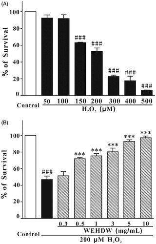
Figure 3. LO2 cells, treated with different agents, under the view of 400 × fluorescence microscope. LO2 cells treated with WEHDW (1 mg/mL) could attenuate apoptotic cell death mediated by H2O2 (200 μM, 6 h).
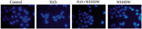
Figure 4. WEHDW attenuated the phosphorylation of MEK and ERK caused by H2O2. (A) PD98059 prevented H2O2-induced cell death in a concentration-dependent manner. After pre-treatment with PD98059 at different concentrations as indicated, LO2 cells were exposed to 200 μM H2O2 for 2 h. Cell viability was measured at 6 h after the H2O2 challenge by MTT assay. Data, expressed as percentage of control, were the mean ± SEM of three separate experiments, ###p < 0.001 versus control and **p < 0.01 versus the H2O2 alone group. (B) H2O2 time-dependently increased the levels of phospho-MEK and phospho-ERK in the first 15 min. LO2 cells were incubated with 200 μM H2O2 at the indicated time points, and total proteins were detected with the use of specific antibodies. WEHDW attenuated the increase of phospho-MEK and phospho-ERK caused by H2O2. LO2 cells were pre-treated with 1 mg/mL WEHDW for 2 h and then exposed to 200 μM H2O2 for 15 min.
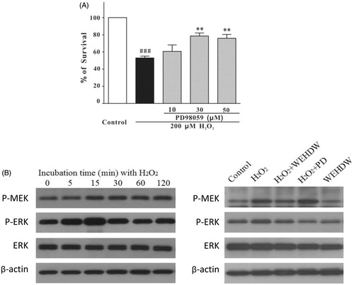
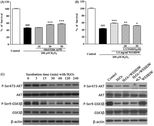
Figure 5. WEHDW activated the phosphorylation of P13-K/AKT/GSK3β pathway. (A) SB415286 prevented H2O2-induced apoptosis in a concentration-dependent manner. LO2 cells were exposed to 200 μM H2O2 at 2 h after pre-treatment with SB415286 at different concentrations as indicated. Cell viability was measured at 6 h after exposure to H2O2 by MTT assay. Data, expressed as percentage of control, were the mean ± SEM of three separate experiments, ###p < 0.001 versus control and ***p < 0.001 versus the H2O2 alone group. (B) Specific P13-K inhibitor abrogated the hepatoprotective effects of WEHDW on H2O2-induced apoptosis. After pre-treatment with 1 mg/mL WEHDW, LO2 cells were incubated with 30 μM or 50 μM LY294002 for 30 min. Then, they were exposed to 200 μM H2O2 at 2 h. Cell viability was measured at 6 h after exposure to H2O2 by MTT assay. Data, expressed as percentage of control, were the mean ± SEM of three separate experiments, ###p < 0.001 versus control and *p < 0.05, **p < 0.01, and ***p < 0.001 versus the H2O2 alone group. (C) H2O2 time-dependently decreased the levels of phospho-AKT and phospho-GSK3β peaked at 30 min without affecting its expression levels. LO2 cells were incubated with 200 μM H2O2 at the indicated time points, and the total proteins were detected with the use of specific antibodies. WEHDW reversed the reduction of phospho-AKT and phospho-GSK3β caused by H2O2. LO2 cells were pre-treated with 1 mg/mL WEHDW for 2 h and then exposed to 200 μM H2O2 for 30 min.