Figures & data
Figure 1. Transmural myocardial infarction (C) in murine left ventricle 12 days after ligation of the LAD. Immunostainings and mRNA analyses were done on cross sections at the periinfarct level (B). Eosin hematoxylin staining. (A) indicates non-infarcted myocardium.
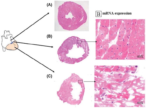
Figure 2. ERα expression in ERαKO, ERβKO and littermate wild type control mice 12 days after induction of myocardial infarction and in non-operated groups. In the infarcted specimens periinfarct tissue was used. Values are mean with SEM.
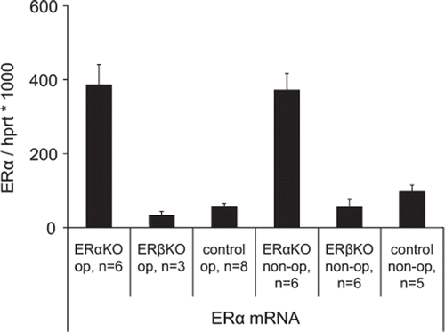
Figure 3. ERβ expression in ERαKO, ERβKO and littermate wild type control mice 12 days after induction of myocardial infarction and in non-operated groups. In the infarcted specimens periinfarct tissue was used. Values are mean with SEM.
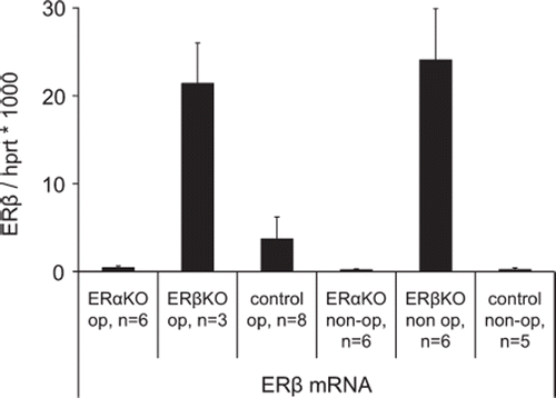
Figure 4. Rabbit polyclonal antibody to mouse was used for detection of ERα in a) positive control, murine brain, b) ERαKO mouse myocardium and c) ERβKO mouse myocardium, (d) littermate control mouse myocardium; and for detection of ERβ in e) positive control, murine brain, f) ERαKO mouse myocardium and g) ERβKO mouse myocardium, (h) littermate control, mouse myocardium. The marker represents 100 μm. Arrows indicate positive cells.
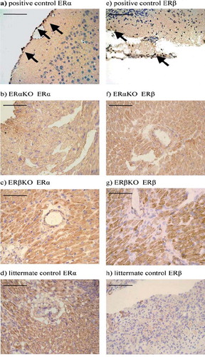
Figure 5. Periinfarct (a) capillary and (b) arteriolar densities 12 days after induction of myocardial infarction in ERαKO, ERβKO, wild type and sham operated mice. While after myocardial infarction vascular densities were lower than in sham operated controls, there were no differences between ERαKO, ERβKO and wild type mice. The assessment was made blinded. Values are mean with SEM.
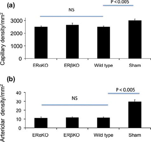
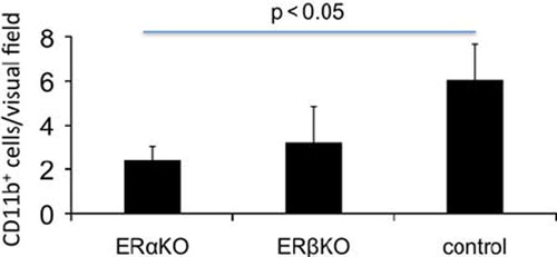
Figure 6. The number of macrophages in the periinfarct area was assessed by counting anti CD11b+ cells. In the periinfarct area of ERαKO mice the number of macrophages was lower compared to control (p < 0.05).