Figures & data
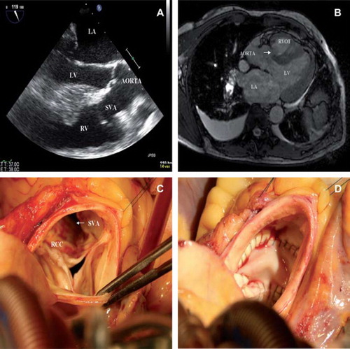
Figure 1. A. Transesophageal echocardiography showing a ruptured sinus of Valsalva (SVA) originating from the right-coronary cusp ending in the right ventricle (RV), left ventricle (LV), left atrium (LA). B. Pre-operative MRI, arrow indicating ruptured sinus of Valsalva aneurysm (SVA). C. Peri-operative picture looking inside the aortic root showing a windsock like aneurismal sac. Right coronary cusp (RCC). D. Peri-operative picture looking inside the aortic root the patch closed defect.