
Figures & data
Figure 1. Schematic presentation of LV filling and ejection spectral Doppler showing how global markers of dyssynchrony, that is, total isovolumic time (t-IVT), Tei index and LV–RV pre-ejection times, are measured. LV: left ventricle; RV: right ventricle; HR: heart rate; ET: ejection time; FT: filling time; IVCT: isovolumic contraction time; IVRT: isovolumic relaxation time.
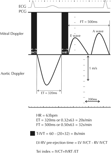
Table I. Clinical and echocardiography data in study patients and in study groups.
Table II. Predictors of CRT non-responders in all patients.
Figure 2. ROC curve showing the highest specificity of combined QRS duration and t-IVT, in predicting CRT non-responders.
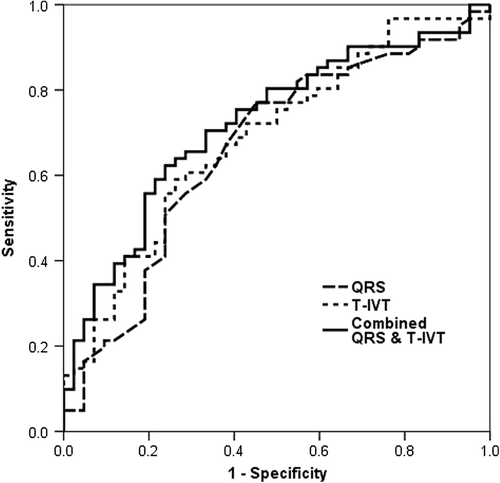
Table III. Predictors of CRT non-responders in atrial fibrillation patients.