Figures & data
Figure 1. Representative ECG and MAP recordings obtained from SAN-driven (panel A) and AV-blocked (panel B) heart preparations. Volume-conducted ECG and RV epicardial MAPs were recorded simultaneously in spontaneously beating heart preparations from two study groups. In panel B (middle trace) the double arrow shows diastolic interval.
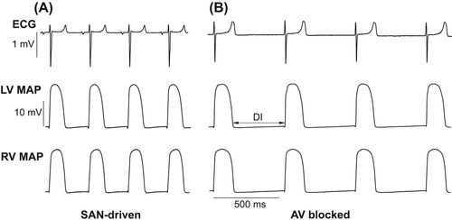
Figure 2. Ventricular APD rate adaptation in SAN-driven (panel A) and AV-blocked (panel B) heart preparations. In panels A and B, sections 1–4 show LV epicardial MAPs obtained while progressively reducing S1–S1 pacing intervals from the maximum value that allowed no escaped beats (section 1) till the minimum value at which 1:1 ventricular capture was preserved (section 4). Further reduction in pacing cycle length resulted in 2:1 conduction block (section 5 in both panels). In all traces, the dashed vertical lines indicate the moments of pacing stimulus application. The S1–S1 pacing interval values are indicated in the first cardiac cycle, and the numbers above MAP traces indicate measured APD90 values. Note that cardiac pacing at S1–S1 =220 ms yields a greater APD90 value in AV-blocked (panel B, section 3) as compared with that in SAN-driven heart preparations (panel A, section 2). Also note a greater value of the minimum pacing interval in AV-blocked (180 ms) as compared with that in SAN-driven heart preparations (110 ms), as shown in section 4 (panels A and B).
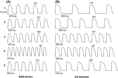
Figure 3. Electrical restitution plots obtained from SAN-driven (panels A, C and E) and AV-blocked (panels B, D, and F) heart preparations. APD90 values determined at three epicardial recording sites in each ventricular chamber were averaged and then plotted as a function of the preceding DI in panels A and B. Panels C and D show changes in APD90 restitution slope over a wide range of diastolic intervals used, and panels E and F show the maximum (Max) restitution slope values determined at the LV and RV epicardium. The dashed rectangular box in panels A and B is to highlight that over a range of diastolic intervals between 49 and 138 ms, the APD90 values determined at similar DIs were longer in AV-blocked as compared with those in SAN-driven heart preparations.
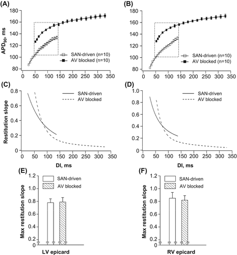
Figure 4. Epicardial APDs (APD90) obtained at identical pacing intervals in SAN-driven and AV-blocked heart preparations. APD90 values determined at three recording sites in LV epicardium (LV epi) and RV epicardium (RV epi) in SAN-driven and AV-blocked heart preparations were averaged and then plotted as a function of S1–S1 pacing interval in panels A and D. Panel B shows individual APD90 values determined at six epicardial recording sites at pacing interval of 260 ms, and panel E shows the relative difference in APD90 value (AV-blocked vs. SAN-driven heart preparations) obtained at each recording site used. The mean APD90 values from 6 recording sites and their SDs are shown in panels C and F, respectively. *P < 0.05 versus its corresponding value in SAN-driven heart preparations (in panels B and C).
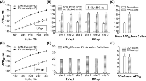
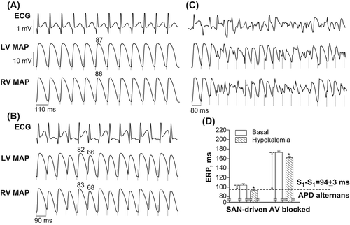
Figure 5. Arrhythmogenic responses during rapid cardiac pacing (panels A, B, and C), and changes in effective refractory periods (panel D) determined in hypokalemic heart preparations. Panels A–C show simultaneous recordings of volume-conducted ECG and LV and RV MAPs obtained upon tachypacing applied to SAN-driven heart preparations subjected to hypokalemic perfusion. In all MAP traces, the dashed vertical lines indicate the moments of pacing stimulus application. The S1–S1 pacing interval values are indicated in the first cardiac cycle (under the RV MAP traces), and the numbers above MAP recordings (panels A and B) indicate measured APD80 values. Panel A: low-amplitude T-wave alternans and regular beat-to-beat APD80 values obtained at S1–S1 = 110 ms. Panel B: shortening of S1–S1 interval till 90 ms provokes high amplitude beat-to-beat oscillations in T wave and APD80. Panel C: further shortening of S1-S1 interval till 80 ms promotes ventricular tachyarrhythmia. Panel D shows that reduced ERPs in hypokalemic setting allow 1:1 capture at S1–S1 intervals producing APD alternans in SAN-driven heart preparations, but not in AV-blocked heart preparations. The double arrows highlight the magnitude of ERP reduction which is required in both groups in order to allow pacing at arrhythmogenic S1–S1 intervals. *P < 0.05 versus its corresponding basal value (in panel D).