Figures & data
Figure 1. Posterolateral view of the left atrium showing CFE distribution before (A) and after (B) flecainide administration. At baseline, CFE areas cover large portions of the lateral and floor segment (A); these are consistently reduced after flecainide administration (B) but substantially preserved in their original localizations. The color scale for different CFE-mean values is displayed on the left (white: < 80 ms and violet: >120 ms). The tracings in the lower panel demonstrate electrograms from the coronary sinus (HALO-5-6) and lateral wall (ROV Abl-D-2) that were collected in the same position (stars) before (A) and after (B) flecainide administration. A significant decrease in deflections detected (yellow spikes) together with a reduction in electrogram amplitude was observed after flecainide administration. CFE, complex fractionated electrogram; LAA, left atrial appendage; LIPV, left inferior pulmonary vein; LSPV, left superior pulmonary vein; MV, mitral valve; RIPV, right inferior pulmonary vein; RSPV, right superior pulmonary vein.
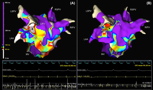
Figure 2. Distribution curves of mapped points sorted by CFE-mean values in mappre (before flecainide, stippled line) and mappost (after flecainide, solid line). Amount of mapped points with relative percentages and statistical differences are presented in the embedded table. CFE, complex fractionated electrogram.
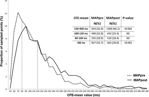
Table I. CFE-mean values in the LA before (mappre) and after (mappost) flecainide administration.
Table II. Distribution of CFE areas by different CFE-mean cutoffs (areas are presented as percentage of the total left atrial surface).
Table III. Size of CFE (CFE mean: < 120 ms) area before (mappre) and after (mappost) flecainide administration.
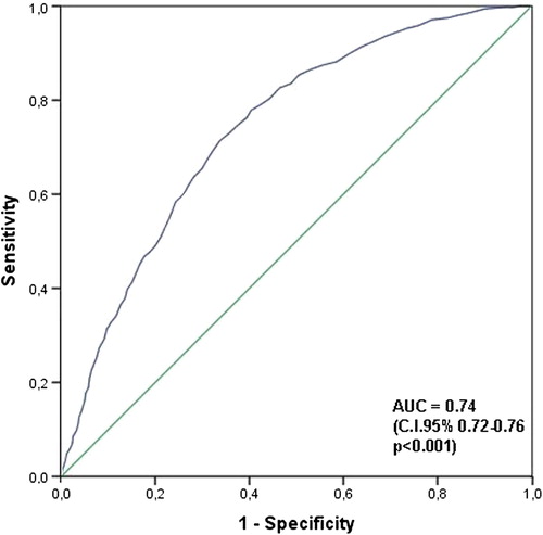
Figure 3. ROC curve: CFE mean in mappre. ROC curve exploring the predictive level of CFE-mean values before flecainide in assessing disappearance of CFE areas after flecainide. AUC, area under the curve; CFE, complex fractionated electrogram.