2,023
Views
97
CrossRef citations to date
0
Altmetric
Research Article

Carbonic anhydrase inhibitors: in vitro inhibition of α isoforms (hCA I, hCA II, bCA III, hCA IV) by flavonoids

, , , &
Pages 283-288 | Received 16 Oct 2011, Accepted 18 Nov 2011, Published online: 14 Dec 2011

Figures & data

Figure 1.  Schematic representation for the three main CA inhibition mechanisms: (A) Sulfonamides (and their isosteres, sulfamate, and sulfamide) substitute the fourth zinc ligand and bind in tetrahedral geometry of the metal ionCitation12; (B) Inorganic anion inhibitors (thiocyanate as an example) add to the metal ion coordination sphere leading to trigonal bipyramidal adductsCitation12; (C) Phenols anchor to the Zn(II) coordinated water molecule/hydroxide ionCitation13; (D) Coumarins (hydrolyzed in situ to 2-hydroxycinnamic acids) occlude the entrance of the active site cavity, interacting both with hydrophilic and hydrophobic amino acid residues. The inhibitor does not interact at all with the catalytically crucial Zn(II) ion which is coordinated by three His residues and a water moleculeCitation10,Citation11.
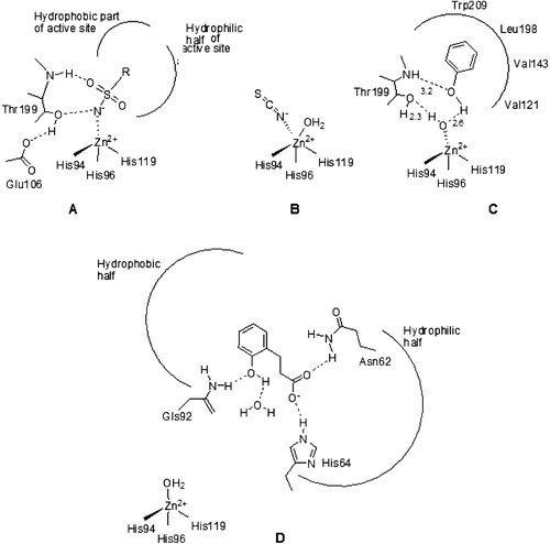
Figure 1.  Schematic representation for the three main CA inhibition mechanisms: (A) Sulfonamides (and their isosteres, sulfamate, and sulfamide) substitute the fourth zinc ligand and bind in tetrahedral geometry of the metal ionCitation12; (B) Inorganic anion inhibitors (thiocyanate as an example) add to the metal ion coordination sphere leading to trigonal bipyramidal adductsCitation12; (C) Phenols anchor to the Zn(II) coordinated water molecule/hydroxide ionCitation13; (D) Coumarins (hydrolyzed in situ to 2-hydroxycinnamic acids) occlude the entrance of the active site cavity, interacting both with hydrophilic and hydrophobic amino acid residues. The inhibitor does not interact at all with the catalytically crucial Zn(II) ion which is coordinated by three His residues and a water moleculeCitation10,Citation11.

Table 1.  KI values (μM) for compound 1–8, EZA, ZNA and AZA of some α-carbonic anhydrase isoforms in human (h) and bovine (b). aRef Citation29.

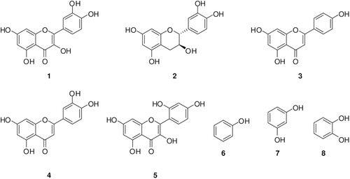
Figure 2.  Structures of common used CA inhİbitors.

Figure 2.  Structures of common used CA inhİbitors.

Figure 3.  Structures of tested compounds 1–8.

Figure 3.  Structures of tested compounds 1–8.

Reprints and Corporate Permissions

Please note: Selecting permissions does not provide access to the full text of the article, please see our help page How do I view content?

To request a reprint or corporate permissions for this article, please click on the relevant link below:

Academic Permissions

Please note: Selecting permissions does not provide access to the full text of the article, please see our help page How do I view content?

Obtain permissions instantly via Rightslink by clicking on the button below:

If you are unable to obtain permissions via Rightslink, please complete and submit this Permissions form. For more information, please visit our Permissions help page.