772
Views
3
CrossRef citations to date
0
Altmetric
Research Article

Thioesters for the in vitro evaluation of agents to image brain cholinesterases

, , , , &
Pages 447-455 | Received 31 Oct 2011, Accepted 29 Nov 2011, Published online: 11 Jan 2012

Figures & data

Table 1.  Affinity constants (Km) and maximum velocity values (Vmax) for N-methylpiperidinyl alkyl thioesters, N-methylpiperidinyl aryl esters and corresponding thioesters with butyrylcholinesterase (BuChE) and acetylcholinesterase (AChE). None of the aryl compounds (1–6) were hydrolyzed by AChE under the conditions used (X).

Figure 1.  Repetitive absorbance scans for N-methylpiperidin-4-yl 4-cyanobenzoate in the presence of butyrylcholinesterase (BuChE) or acetylcholinesterase (AChE). Note change in absorbance over time when the compound was incubated with BuChE (left) reflecting hydrolysis of the compound by this enzyme. No change in absorbance with AChE (right) indicates no hydrolysis by this enzyme. The absorbance was measured every 2 min for 30 min.
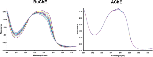
Figure 1.  Repetitive absorbance scans for N-methylpiperidin-4-yl 4-cyanobenzoate in the presence of butyrylcholinesterase (BuChE) or acetylcholinesterase (AChE). Note change in absorbance over time when the compound was incubated with BuChE (left) reflecting hydrolysis of the compound by this enzyme. No change in absorbance with AChE (right) indicates no hydrolysis by this enzyme. The absorbance was measured every 2 min for 30 min.

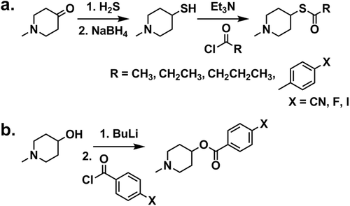
Scheme 1.  Reaction scheme for the synthesis of (a) alkyl and aryl thioesters and (b) aryl esters.

Scheme 1.  Reaction scheme for the synthesis of (a) alkyl and aryl thioesters and (b) aryl esters.

Figure 2.  Histochemical staining of human brain tissue at the level of the thalamus in the coronal plane. (A) Parcellation of the thalamus in the region used to compare histochemical staining by various cholinesterase substrates; (B) Acetylthiocholine. (C) (N-methylpiperidin-4-yl) ethanethioate. Note that both of these substrates produced a similar pattern of staining recapitulating the known distribution of acetylcholinesterase in this region. (D) Butyrylthiocholine. (E) (N-methylpiperidin-4-yl) butanethioate. (F) (N-methylpiperidin-4-yl) 4-cyanobenzenecarbothioate. Note that these substrates produced a similar pattern of distribution that reflected the known distribution of butyrylcholinesterase in this region. AV: anteroventral nucleus; CL: central lateral nucleus; MD: mediodorsal nucleus; R: reticular nucleus; V: ventral nuclei. Scale bar = 200 µm.
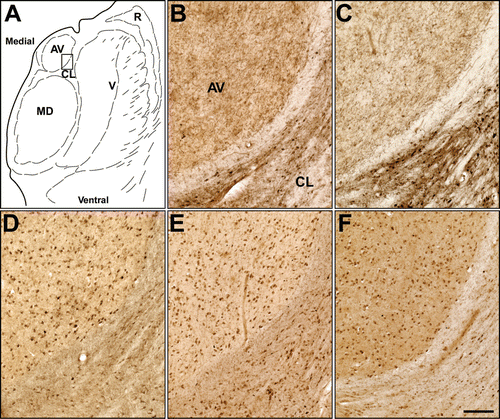
Figure 2.  Histochemical staining of human brain tissue at the level of the thalamus in the coronal plane. (A) Parcellation of the thalamus in the region used to compare histochemical staining by various cholinesterase substrates; (B) Acetylthiocholine. (C) (N-methylpiperidin-4-yl) ethanethioate. Note that both of these substrates produced a similar pattern of staining recapitulating the known distribution of acetylcholinesterase in this region. (D) Butyrylthiocholine. (E) (N-methylpiperidin-4-yl) butanethioate. (F) (N-methylpiperidin-4-yl) 4-cyanobenzenecarbothioate. Note that these substrates produced a similar pattern of distribution that reflected the known distribution of butyrylcholinesterase in this region. AV: anteroventral nucleus; CL: central lateral nucleus; MD: mediodorsal nucleus; R: reticular nucleus; V: ventral nuclei. Scale bar = 200 µm.
Supplemental material

Supplementary Material

Download PDF (216.2 KB)

Reprints and Corporate Permissions

Please note: Selecting permissions does not provide access to the full text of the article, please see our help page How do I view content?

To request a reprint or corporate permissions for this article, please click on the relevant link below:

Academic Permissions

Please note: Selecting permissions does not provide access to the full text of the article, please see our help page How do I view content?

Obtain permissions instantly via Rightslink by clicking on the button below:

If you are unable to obtain permissions via Rightslink, please complete and submit this Permissions form. For more information, please visit our Permissions help page.