829
Views
23
CrossRef citations to date
0
Altmetric
Research Article

Evaluation of a dithiocarbamate derivative as an inhibitor of human glutaredoxin-1

, , , &
Pages 456-462 | Received 30 Sep 2011, Accepted 08 Dec 2011, Published online: 03 Feb 2012

Figures & data

Figure 1.  Mechanism of catalysis of deglutathionylation by GRX. A glutathionylated protein (PS-SG) is restored by the action of RX. GRX operates by a monothiol mechanism in the process of deglutathionylation. The glutathione (GSH) is transferred from the protein to GRX. A GSH molecule restores the normal GRX active site structure with the production of GSSG. The GSSG produced is reduced to GSH by the action of GR which uses NADPH as a cofactor.

Figure 1.  Mechanism of catalysis of deglutathionylation by GRX. A glutathionylated protein (PS-SG) is restored by the action of RX. GRX operates by a monothiol mechanism in the process of deglutathionylation. The glutathione (GSH) is transferred from the protein to GRX. A GSH molecule restores the normal GRX active site structure with the production of GSSG. The GSSG produced is reduced to GSH by the action of GR which uses NADPH as a cofactor.

Figure 2.  Structure of 2-AAPA.

Figure 2.  Structure of 2-AAPA.

Figure 3.  Time and concentration dependence of GRX-1 inhibition by 2-AAPA. The natural logarithm of GRX-1 remaining activity is plotted against time. The enzyme was incubated with increasing concentrations of 2-AAPA, and aliquots were withdrawn at various time points for determination of remaining GRX-1 activity. The graph shows a representative plot from one of triplicate experiments. ▪, 200 μM; ▴, 100 μM; ×, 50 μM; •, 25 μM.
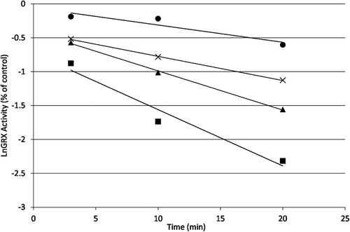
Figure 3.  Time and concentration dependence of GRX-1 inhibition by 2-AAPA. The natural logarithm of GRX-1 remaining activity is plotted against time. The enzyme was incubated with increasing concentrations of 2-AAPA, and aliquots were withdrawn at various time points for determination of remaining GRX-1 activity. The graph shows a representative plot from one of triplicate experiments. ▪, 200 μM; ▴, 100 μM; ×, 50 μM; •, 25 μM.

Figure 4.  Double reciprocal plot. The reciprocals of the apparent rate constants of inhibition (slopes from ) are plotted against the reciprocals of 2-AAPA concentration. The graph shows a representative plot from one of triplicate experiments.
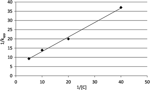
Figure 4.  Double reciprocal plot. The reciprocals of the apparent rate constants of inhibition (slopes from Figure 3) are plotted against the reciprocals of 2-AAPA concentration. The graph shows a representative plot from one of triplicate experiments.

Figure 5.  Irreversibility of GRX-1 inhibition by 2-AAPA. GRX-1 was incubated with 2-AAPA 1 mM for 1 hour to achieve complete inhibition. Then the solution was transferred to a DisPoDialyzer and dialyzed in Tris buffer for 4 hours. No enzyme activity returned in the 2-AAPA sample over a 4 hour period. The graph shows a representative plot from one of triplicate experiments. ♦, control; ▪, 2-AAPA treated.
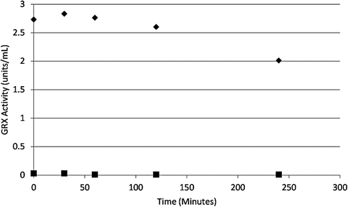
Figure 5.  Irreversibility of GRX-1 inhibition by 2-AAPA. GRX-1 was incubated with 2-AAPA 1 mM for 1 hour to achieve complete inhibition. Then the solution was transferred to a DisPoDialyzer and dialyzed in Tris buffer for 4 hours. No enzyme activity returned in the 2-AAPA sample over a 4 hour period. The graph shows a representative plot from one of triplicate experiments. ♦, control; ▪, 2-AAPA treated.

Figure 6.  Substrate protection of GRX from 2-AAPA inhibition. GRX substrate GSH-HED protected the enzyme from inhibition by 2-AAPA in a concentration dependent manner indicating that 2-AAPA is a competitive inhibitor of GRX-1 (n = 3).
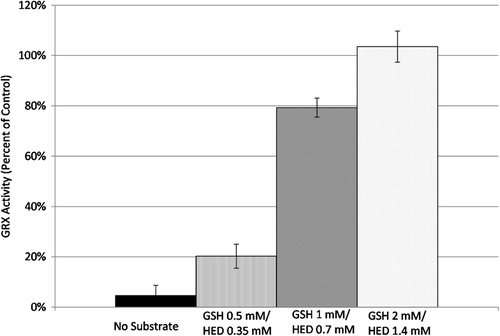
Figure 6.  Substrate protection of GRX from 2-AAPA inhibition. GRX substrate GSH-HED protected the enzyme from inhibition by 2-AAPA in a concentration dependent manner indicating that 2-AAPA is a competitive inhibitor of GRX-1 (n = 3).

Figure 7.  (A) LC/MS analysis of covalent binding of 2-AAPA to GRX-1. GRX-1 inhibited by 2-AAPA 0.1 mM for 20 minutes. The native enzyme has an m/z of 11641. In the inhibited sample, additional peaks are observed at m/z 11,997 and 12,353, corresponding to monothiocarbamoylation at one or two cysteines, respectively. (B) LC/MS analysis of covalent binding of 2-AAPA to GRX. GRX-1 with 2-AAPA 0.1 mM and substrate (GSH 2 mM/HED 1.4 mM). The signals from the addition of 2-AAPA are not observed in the sample incubated with inhibitor and substrate; the signal at m/z 11,948 corresponds to the addition of glutathione.
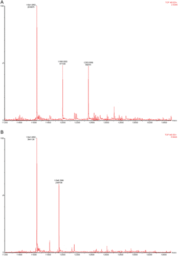
Figure 7.  (A) LC/MS analysis of covalent binding of 2-AAPA to GRX-1. GRX-1 inhibited by 2-AAPA 0.1 mM for 20 minutes. The native enzyme has an m/z of 11641. In the inhibited sample, additional peaks are observed at m/z 11,997 and 12,353, corresponding to monothiocarbamoylation at one or two cysteines, respectively. (B) LC/MS analysis of covalent binding of 2-AAPA to GRX. GRX-1 with 2-AAPA 0.1 mM and substrate (GSH 2 mM/HED 1.4 mM). The signals from the addition of 2-AAPA are not observed in the sample incubated with inhibitor and substrate; the signal at m/z 11,948 corresponds to the addition of glutathione.

Figure 8.  Proposed mechanism of 2-AAPA binding to GRX-1. Data from mass spectrometry analysis of the inhibited enzyme show that 2-AAPA binds covalently to GRX-1. Monothiocarbamoylation of the enzyme can occur at one or two cysteines in GRX. Based on the pKa, it is likely that the monothiocarbamoylation leading to GRX-1 inhibition is occurring at CYS-22.

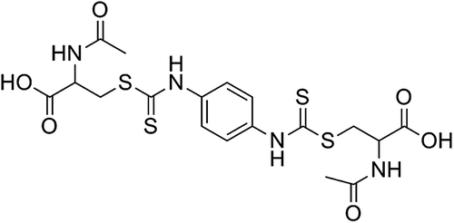
Figure 8.  Proposed mechanism of 2-AAPA binding to GRX-1. Data from mass spectrometry analysis of the inhibited enzyme show that 2-AAPA binds covalently to GRX-1. Monothiocarbamoylation of the enzyme can occur at one or two cysteines in GRX. Based on the pKa, it is likely that the monothiocarbamoylation leading to GRX-1 inhibition is occurring at CYS-22.

Reprints and Corporate Permissions

Please note: Selecting permissions does not provide access to the full text of the article, please see our help page How do I view content?

To request a reprint or corporate permissions for this article, please click on the relevant link below:

Academic Permissions

Please note: Selecting permissions does not provide access to the full text of the article, please see our help page How do I view content?

Obtain permissions instantly via Rightslink by clicking on the button below:

If you are unable to obtain permissions via Rightslink, please complete and submit this Permissions form. For more information, please visit our Permissions help page.