Figures & data
Figure 1. Left panel: 2D sketch of the binding mode of Clorobiocin into its binding site of DNA gyrase-B, showing three hydrogen bonds as green dotted lines (with residues Arg136, Asp73 and Asn46) and one Pi-cationic interaction with Arg76; right panel: Docking validation of Clorobiocin with DNA gyrase-B: crystal structure ligand (red) docked ligand (yellow); RMSD 0.41 Å.
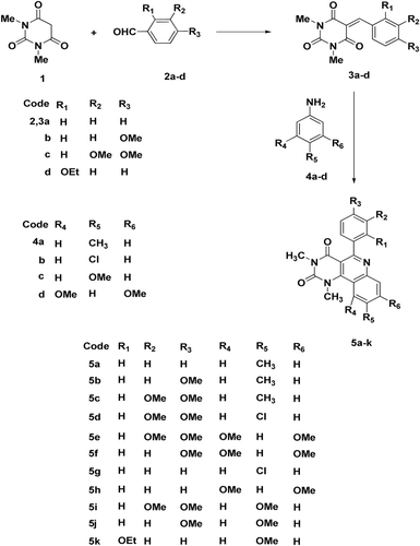
Figure 2. Synthesis of pyrimido[5,4-c]quinoline-2,4-dione derivatives 5a–k.
![Figure 2. Synthesis of pyrimido[5,4-c]quinoline-2,4-dione derivatives 5a–k.](/cms/asset/654390cd-59a3-45f9-beac-bc1b6703ddac/ienz_a_654113_f0002_b.gif)
Table 1. In vitro antimicrobial activity of pyrimido[5,4-c]quinoline-2,4-diones 5a–k.
Figure 3. 2D Ligand interaction of the 5-aryl-pyrimido[5,4-c]quinoline-2,4-diones with DNA gyrase-B; panel (1) compound 5c showing only pi-cationic interaction with Arg76; panel (2) compound 5e showing pi-cationic interaction with Arg76 and H-bond with Arg136; panel (3) compound 5f showing pi-cationic interaction with Arg76 and two H-bonds with Arg136 and Asn46.
![Figure 3. 2D Ligand interaction of the 5-aryl-pyrimido[5,4-c]quinoline-2,4-diones with DNA gyrase-B; panel (1) compound 5c showing only pi-cationic interaction with Arg76; panel (2) compound 5e showing pi-cationic interaction with Arg76 and H-bond with Arg136; panel (3) compound 5f showing pi-cationic interaction with Arg76 and two H-bonds with Arg136 and Asn46.](/cms/asset/75a2483f-6a01-4217-88a4-648f66e13f16/ienz_a_654113_f0003_b.gif)
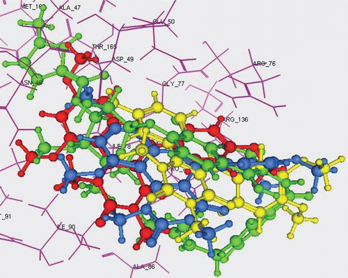
Figure 4. 3D overlay of Clorobiocin (green), 5c (yellow), 5e (blue) and 5f (red) in DNA-gyrase active pocket.