Figures & data
Table 1. Distribution of CA III in different mouse tissues.
Figure 1. Western blot of recombinant human CA III identified with the new anti-CA III antibody. The antibody identifies a single 30-kDa polypeptide band. No reactions are seen with the other isozymes.
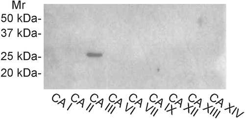
Figure 2. Western blotting of mouse tissues. The strongest 30-kDa polypeptide bands of CA III are detected in the liver, soleus and EDL muscles. Moderate or very faint bands are seen in the stomach, ileum, colon, submandibular gland, kidney, testis, epididymis, uterus, heart, lung, and cerebellum. Female (above) and male (below) mouse tissues were investigated separately.
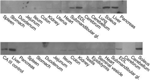
Figure 3. Immunohistochemical staining of CA III in different mouse tissues. The EDL muscle (a) shows occasional strongly stained muscle fibers, while a weaker positive signal is observed in the heart (b). The kidney (c) shows only very faint staining in the renal tubules. In the testis (d), a very weak positive signal is detected in the interstitial cells (arrow). The lung (e) and spleen (f) are completely negative. Bars 50 µm.
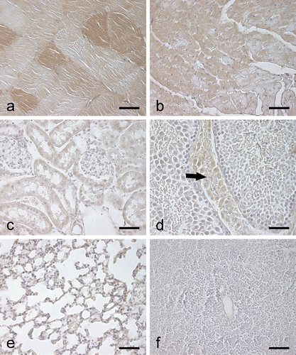
Figure 4. Immunohistochemical staining of CA III in mouse gastrointestinal organs. In the liver (a), the most intense staining is observed in the hepatocytes of perivenular region (cv, central vein). Control staining using pre-immune rabbit serum is negative except for nonspecific nuclear reactions (b). In the submandibular gland (c), the epithelial cells of the salivary duct (arrows) show very weak immunostaining. The stomach also shows a weak positive signal (d), while no staining is detected in the small intestine (e) and colon (f). Bars 50 µm.
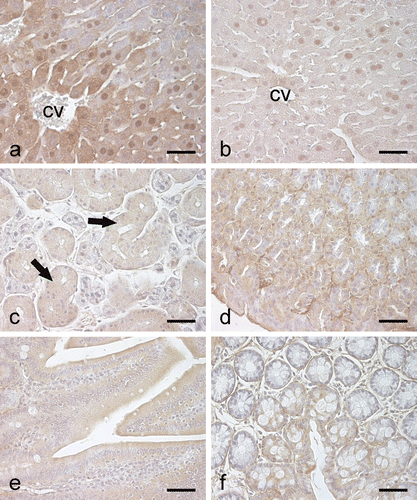
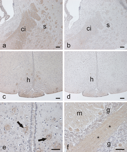
Figure 5. Immunostaining of CA III in the mouse brain. In the cerebrum, the capsula interna (a) shows moderate positive reaction, while the control staining is negative (b). (ci, capsula interna; s, striatum). Some neurons in the hypothalamic region are stained (c) (h, hypothalamus). Control shows no reaction (d). (e) A higher-magnification image in which the CA III-positive staining in the hypothalamic neurons (arrows) is seen more clearly. In the cerebellum (f), some neuronal axons and the Purkinje cells are positively stained. (m, molecular layer; g, granule cell layer; *, neuronal axons). Bars 50 µm.