Figures & data
Table 1. Molecular properties of human and mouse CARPs.
Table 2. Distribution of CARP VIII protein and its mRNA in brain and other tissues.
Figure 1. Expression pattern of CA8 gene in human analyzed by microarray method. The expression profile figure is adapted from BioGPS (http://biogps.gnf.org, accessed July 2012). The figures shows expression of CA8 gene in different human tissues samples and expression is significantly high in cerebellum compared to other tissuesCitation20.
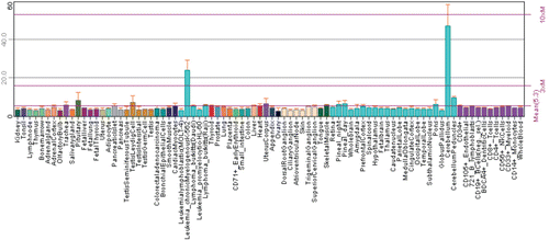
Figure 2. Expression of CA8 gene in normal and pathological conditions in human. Microarray data of the mRNA expression levels of human genes in normal and pathological tissues from MediSapiens. (http://www.medisapiens.com, accessed February 2012)Citation21.
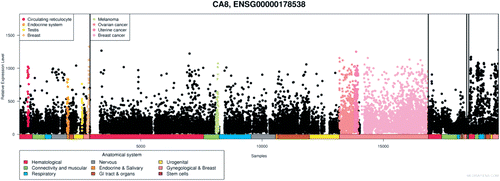
Figure 3. Reduction in cerebellar volume of Saudi Arabian patients with a mutation in CA8 gene (Gln162-Arg). The brain magnetic resonance imaging (MRI) of the (A–D) patient 1, (E–H) patient 2, and the (I–L) patient 3. Figures show the progressive cerebellar loss in patient 1 between the sagittal T1 (A and C) and axial flair images (B and D) in the scans performed at 4 (A and B) and 8 years of age (C, D), respectively. Sagittal T1 images (E and I) show reduction in cerebellar volume more prominent in patient 3 (I) than in patient 2 (E). The volume loss is not associated with parenchymal signal abnormality as evident in images F, G, J, and K. Small sella turcica and skull base fatty replacement are also shown (E and I). Axial flair images (H and L) demonstrate deep periventricular white matter abnormalities with more involvement seen in patient 3 (L). Modified from Kaya et al.Citation7
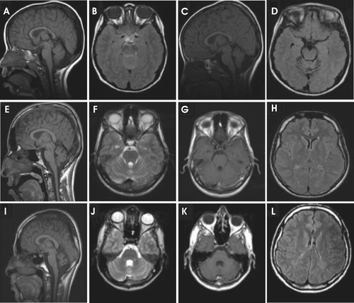
Figure 4. Locations of the CA8 gene variation in members of Iraqi and Saudi Arabian family. (A) The location of the variation (yellow arrow, Glycine 162 Arginine) on hypothetical 3-D structure of carbonic anhydrase-related protein VIII (CARP VIII) protein as shown with incorrect numbering in Kaya et al.Citation7 (B) The 3-D structure of human CARP VIII (PDB ID: 2W2J) showing the correct places of variations in the CARP VIII protein (substitution of amino acids) found in members of Iraqi (top right black arrow, Serine 100 Proline) and members of Saudi Arabian family (bottom right black arrow Glycine 162 Arginine), top left black arrow, the position of panel A correctly identified as Glycine 193.
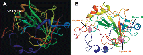
Table 3. Distribution of CARP X protein and its mRNA in brain and other tissues.
Figure 5. Expression profile of Car10 mRNA in murine tissues. The relative expression pattern of Car8 mRNAs in 20 different mouse tissues using RT-qPCR. The predominant expression of Car8 mRNA is seen central nervous system (CNS), expression being very high in cerebellum followed by parietal cortex and frontal cortex, very low level of expression was also seen in midbrain and eye (). Adopted from Aspatwar et al.Citation12
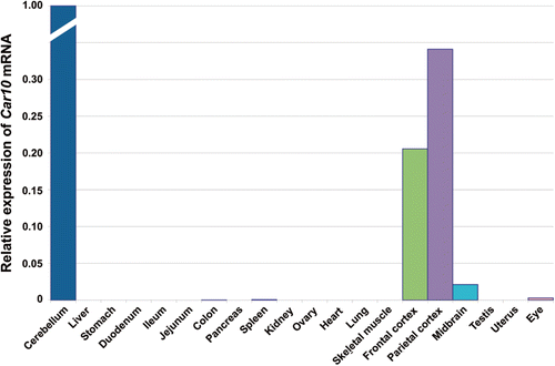
Figure 6. Immunohistochemical staining of carbonic anhydrase-related protein X (CARP X) protein in mouse tissues. Panel I. Expression of CARP X in the brain. (A) There was no expression in cerebellum. (B) The arrowheads in figure show CARP X-positive microcapillaries in the cerebrum. Panel II. Moderate CARP X expression was observed in the respiratory epithelium (arrowhead) of the lung (G). Moderate expression was also present in the gastric glands (arrows in panel D). The heart muscle cells occasionally showed extremely weak signals (I). The other tissues including the (A) liver, (B) pancreas, (C) submandibular gland, (E) colon, (F) small intestine, (H) skeletal muscle remained negative. Original magnifications are at ×20. Adopted from Aspatwar et al.Citation12
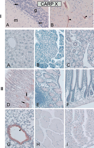
Figure 7. Expression pattern of CA10 gene in healthy human tissues analyzed by microarray method. The expression profile figure is adapted from BioGPS (http://biogps.gnf.org, accessed July 2012). The figures shows expression of CA10 gene in different human tissues samples and expression is markedly high in the pineal gland during the night compared to day time. Some expression can also be seen in cerebellum and other parts of the brain.Citation20
Figure 8. Expression of CA10 gene in normal and pathological conditions in human: Microarray data of the mRNA expression levels of human genes in normal and pathological tissues from MediSapiens. (http://www.medisapiens.com, accessed February 2012)Citation21.
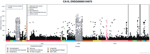
Table 4. Distribution of CARP XI protein and its mRNA in brain and other tissues.
Figure 9. Distribution of Car11 mRNA in murine tissues. The relative expression pattern of Car11 mRNAs in 20 different mouse tissues analyzed using reverse transcription-quantitative PCR (RT-qPCR). The overall pattern of Car11 mRNA expression was similar to the expression of Car8 mRNA ( and ). The expression of mRNA and can be seen in most of the tissues. The Car11 mRNA was predominantly expressed in frontal cortex followed by significantly high level of expression in parietal cortex, cerebellum and midbrain. Low amount of Car11 mRNA signal was also seen in colon, kidney, ovary lung and eye (). Adopted from Aspatwar et al.Citation12
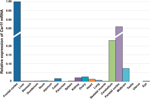
Figure 10. Immunohistochemical staining of carbonic anhydrase-related protein XI (CARP XI) protein in mouse tissues. Panel I. (A) Moderate expression of CARP XI in cerebellar Purkinje cells. (B) There was faint signal for CARP XI in cerebrum. Panel II. Weak immunoreactions for CARP XI in the crypts of Lieberkuhn (arrows in D), gastric glands (arrows in C), and renal tubule cells (F). Extremely weak signals can also be observed in the (A) liver, (E) colon and (I) testis. The (B) pancreas, (G) lung, and (H) skeletal muscle were all negative. Original magnifications are at ×20. Adopted from Aspatwar et al.Citation12
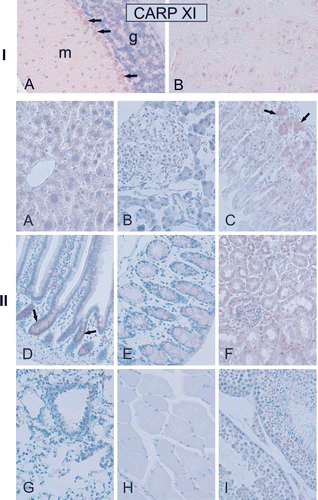
Figure 11. Expression pattern of CA11 gene in healthy human tissues analyzed by microarray method. The expression profile figure is adapted from BioGPS (http://biogps.gnf.org, accessed July 2012). The figures shows expression of CA11 gene in different human tissues samples and significant level of expression can only be seen in central nervous systemCitation20.
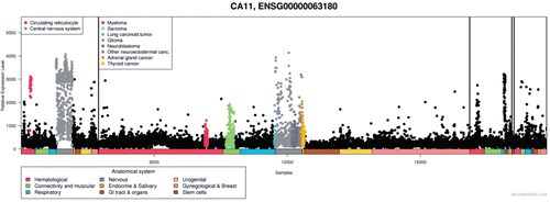
Figure 12. Expression of CA11 gene in normal and pathological conditions in human: Microarray data of the mRNA expression levels of human genes in normal and pathological tissues from MediSapiens. (http://www.medisapiens.com, accessed February 2012)Citation21.