Figures & data
Figure 1. Chemical structures of doxorubicin, melphalan and acetazolamide.
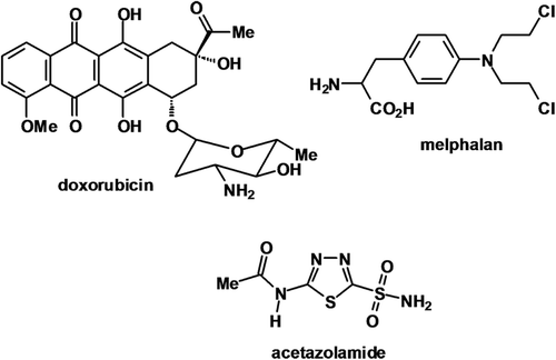
Table 1. Inhibition of CA IX activity by AZM.
Figure 2. CA activity of membrane extracts in air and anoxia. (A) Wide range of aerobic (black bars) and anoxic (white bars) levels of CA activity observed, for example HCT116 and EV1 cells exhibited lower activity levels with minimal anoxic induction. CA9/18 cells had high aerobic activity and moderate induction under anoxic conditions. Activity in HT29 cells was greatly enhanced under anoxic conditions. (B) CA IX protein expression in the four cell lines specified reflected the activity profiles. (C) 10–100 µM AZM reduced activity in EV1 (black line) and CA9/18 (grey line) cells in a dose dependent manner under aerobic conditions. *p < 0.05. Error bars indicate SEM of n = 3 samples. AZM, acetazolamide; CA IX, carbonic anhydrase IX.
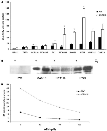
Figure 3. AZM reduces toxicity of melphalan. AZM alone had a minor impact on cell survival, but the results were corrected for any effects of AZM. (A) 50 µM AZM reduced MEL toxicity in aerobic (left panel) and anoxic (right panel) HT29 cells. (B) No reduction in MEL toxicity following 50 µM AZM exposure under aerobic conditions was noted in HCT116 (black bars) and EV1 (white bars) cells (left panel). 50 µM AZM significantly reduced 50 µM MEL toxicity in HT29 cells (6.3 fold) under anoxic conditions (horizontal lined bars) (right panel). *p < 0.05. Error bars indicate SEM of n = 3 samples. AZM, acetazolamide; MEL, melphalan.
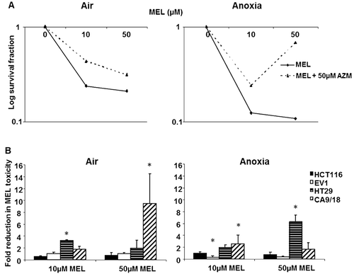
Figure 4. AZM enhances toxicity of doxorubicin. The minimal effect of AZM alone on toxicity has been corrected. (A) The addition of 50 µM AZM increased DOX toxicity in HT29 cells under aerobic and anoxic conditions. (B) 50 µM AZM had a minimal effect on doxorubicin toxicity in HCT116 (black bars) and EV1 (white bars) cells under aerobic conditions (left panel), however CA9/18 (diagonal lined bars) cells showed a 5.2 fold increase in toxicity of 5 µM DOX. 50 µM AZM did not alter DOX toxicity in HCT116 and EV1 cells under anoxic conditions (right panel), but HT29 (horizontal lined bars) cells exhibited an approximately 5 fold increase in toxicity at both doses of DOX. Anoxic CA9/18 cells showed a 5.2 fold increase in toxicity of 1 µM DOX and 12.13 fold increase of 5 µM DOX toxicity (right panel). *p < 0.05. Error bars indicate SEM of n = 3 samples. AZM, acetazolamide; DOX, doxorubicin.
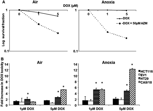
Figure 5. AZM limited GSH depletion by MEL but enhanced uptake of doxorubicin. (A) Under aerobic conditions no cell line revealed significant increased GSH levels upon the addition of 50 µM AZM to MEL (left panel). Under anoxic conditions, HT29 cells (horizontal lined bars) showed a 5.33 fold increase in GSH levels at 50 µM MEL in combination with 50 µM AZM, but other cell lines were unaffected (right panel). (B) AZM alone had no effect on background fluorescence in the cell line tested in air or anoxia. DOX increased the mean fluorescence value relative to concentration (data not shown), but was observed to be lower in anoxia than normoxia. Fluorescence was unaffected in all the cell lines by the addition of 50 µM AZM under aerobic conditions (left panel). Under anoxic conditions, 50 µM AZM significantly enhanced the uptake of 1 µM DOX in HT29 cells (horizontal lined bars) and induced a 12 fold increase in the toxicity of 5 µM DOX in CA9/18 cells (diagonal lined bars) (right panel). *p < 0.05 Error bars indicate SEM of n = 3 samples. AZM, acetazolamide; DOX, doxorubicin; GSH, glutathione; MEL, melphalan.
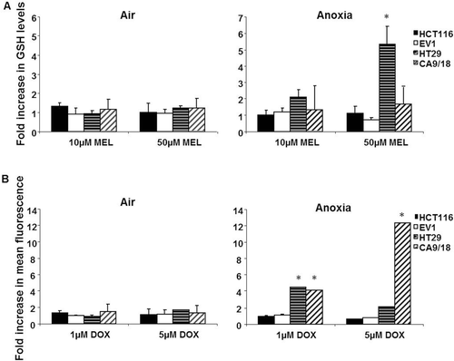
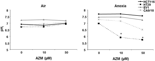
Figure 6. AZM reduced intracellular pH (pHi). The pHi of all the cell lines tested was around neutral and was not substantially affected by AZM under aerobic conditions (left panel). Under anoxic conditions the control pHi (0 µM AZM) in all lines slightly increased in comparison with air. HCT116 and EV1 pHi remained unaltered by AZM treatment under anoxic conditions (solid lines). CA9/18 cells (grey dashed line) exhibited decreased pHi at 50 µM AZM in anoxia. The pHi of anoxic HT29 cells significantly reduced with both concentrations of AZM (right panel). *p < 0.05. Error bars indicate SEM of n = 3 samples. AZM, acetazolamide.