Figures & data
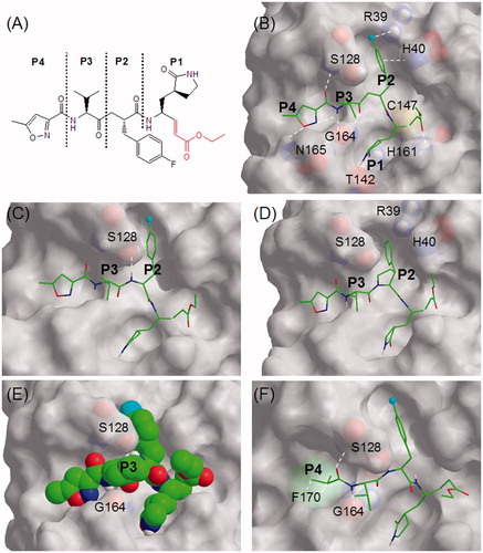
Figure 1. (A) Schematic diagram of Rupintrivir showing P1, P2, P3, and P4 regions using conventional terminologyCitation11. The ethyl propenoate moiety is depicted in red; (B) the stick model of Rupintrivir (green) in the EV71 3C protease catalytic site (3SJO.pdb). Protease residues involved in ligand interaction are numbered and shown as CPK representations, the cyan sphere represents fluorine and white hashes represent possible interactions; (C) Cpd. 2 showing the extra H-bond between the P2 NH and S128; (D) Cpd. 9 with its P2 phenylpyrrolidine moiety oriented in the S2 subsite; (E) the CPK model of Cpd. 16 showing its P3 cyclohexyl sandwiched between S128 and G164; (F) Cpd. 21 showing hydrophobic interactions between its P4 N-cap and F170 phenyl group (green CPK representation) in the S4 subsite. Figures C–F were obtained from molecular modeling.
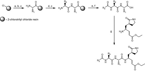
Scheme 1. General synthetic procedure for compounds 2–31. Reagents and conditions: (a) swell 2-chlorotrityl chloride resin in DMF, 25 °C, 30 min; (b) appropriate Fmoc-protected amino acid: Fmoc-R1-OH, DIPEA, CH2Cl2, 25 °C, 30 min; (c) 20% piperidine in DMF (v/v), 25 °C, 30 min; (d) appropriate Fmoc-protected amino acid: Fmoc-R2-OH, HBTU, HOBT, DIPEA, DMF, 25 °C, 1 h; (e) appropriate carboxylic acid: R3COOH, HBTU, HOBt, DIPEA, DMF, 25 °C, 1 h; (f) 95% TFA in CH2Cl2 (v/v), 25 °C, 30 min; (g) (S,E)-ethyl 4-amino-5-(S-2-oxopyrrolidin-3-yl)pent-2-enoate, DIC, HOBT, DMF, 25 °C, 16 h.