Figures & data
Figure 1. Chemical structure of previously reported pyrazolo[3,4-d]pyrimidines endowed with anti-cancer and apoptosis inducing activities (A–D) and the synthesized compounds (E).
![Figure 1. Chemical structure of previously reported pyrazolo[3,4-d]pyrimidines endowed with anti-cancer and apoptosis inducing activities (A–D) and the synthesized compounds (E).](/cms/asset/a2ab83b3-d4ab-4630-9c0e-be98963c0602/ienz_a_1118686_f0001_c.jpg)
Scheme 1. Synthetic pathways for the target compounds 5–7.

Scheme 2. Synthetic pathways for the target compounds 9–11.
Figure 2. Compounds 7a and 7b reduced cell viability and induced apoptosis in RKO cells. (A) A dose–response curve for compounds 7a and 7b on FHC normal colon cancer cells was constructed. Each data point was an average of results from three independent experiments performed in triplicate and presented as mean ± SD. (B) Comparison of 7a and 7b cytotoxicity in FHC and RKO cells. Both cells were treated with 8 and 4 μM of compounds 7a and 7b, respectively. Untreated cells were used as control. (C) Comparison of 7a and 7b induced apoptosis in FHC and RKO cells. FHC and RKO cells were treated with 8 and 4 μM of compounds 7a and 7b, respectively, for 24 h. Untreated cells were used as control. ELISA assay was applied for apoptotic cell detection. Each experiment was performed in triplicate. Data are presented as mean ± SD (n = 3). Significant differences between the control and test compounds are indicated by * (*p < 0.05). (D) TUNEL assay was used to confirm induction of apoptosis in RKO cells. Cells were treated with 8 and 4 μM of 7a and 7b, respectively, for 24 h. Untreated cells act as control. Lack of staining in control (untreated) cells, represents that the cells are actively proliferating, i.e. without apoptotic cell death and induction of apoptosis in treated cells was confirmed by appearance of TUNEL positive cells. Images were acquired for each fluorescence channel, using suitable filters at 200 × magnification.
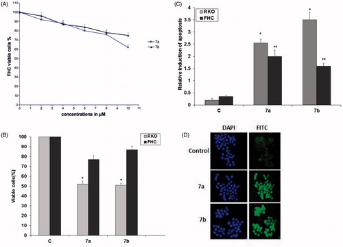
Table 1. IC50 (μM)Table Footnote* of the synthesized 6-substituted 1H-pyrazolo[3,4-d]pyrimidin-4(5H)-ones against five cancer cell lines.

Figure 4. Effect of compound 7b on apoptotic signaling in RKO cells. (A) Cells were treated with 4 μM of compound 7b for 24 h. The cell lysates were collected and the levels of p53, Akt, Bcl-2, ERK1/2 were studied by Western blotting analysis using specific antibodies. β-Actin was used as loading control. C: control (untreated cells). (B) Total protein and mitochondrial fraction were exacted and Western blotting analysis was applied to determine the protein levels of Bax in treated and non-treated cells. β-Actin was used as loading control and IV-Cox was used as internal mitochondrial control.
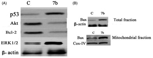
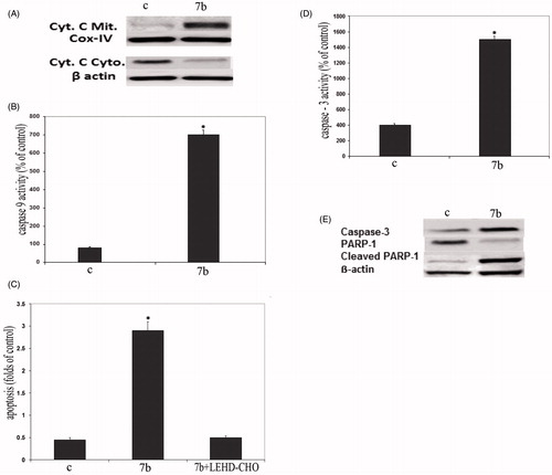
Figure 5. Compound 7b induced apoptosis through initiation of the mitochondrial pathway. (A) Western blot analysis was used to determine the release of cytochrome c in the cytosol and mitochondria of 7b-treated RKO cells. β-Actin was used as loading control and IV-Cox was used as internal mitochondrial control. C: control (untreated cells). (B) Effect of 7b on the level of caspase-9 in RKO cells after treatment with 4 μM 7b for 24 h. (C) Effect of 7b and its combination with caspase-9 inhibitor on 7b-induced apoptosis. Cells were preincubated with 20 μM LEHD-CHO for 1 h before the addition of 4 μM of 7b for an additional 24 h. Induction of apoptosis was determined at 405 nm and compared to control (untreated cells). (D) Activation of caspase-3 in RKO cells after treatment with 4 μM 7b for 24 h. (For B, C and D; each data point is the mean of three independent experiments and expressed as M ± SD. The asterisk indicates a significant difference between control (C; untreated cells) and 7b-treated cells; p < 0.05). (E) Western blot analysis was used to determine the expression levels of caspase-3 and cleaved PARP1 after treating cells with 7b for 24 h. β-Actin was used as loading control.