Figures & data
Figure 1. Histopathological examination of the placenta from a previous pregnancy showed fibrinoid deposition (arrow) in the intervillous space surrounding more than 50% of the villi in some full-thickness sections (H&E; 40×) (a) and absence of physiologic transformation of a spiral artery, i.e. persistent muscularization (circle) in the basal plate (H&E, 100×) (b).
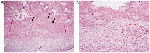
Figure 2. Maternal plasma concentrations (log base e) of soluble vascular endothelial growth factor receptor -1 (sVEGFR-1) (a), placental growth factor (PlGF) (b), soluble endoglin (sEng) (c) and the ratio of PlGF/sVEGFR-1 (d) throughout pregnancy plotted against reference ranges at 2.5th 5th, 10th, 50th, 90th, 95th, and 97.5th percentile of uncomplicated pregnancies.
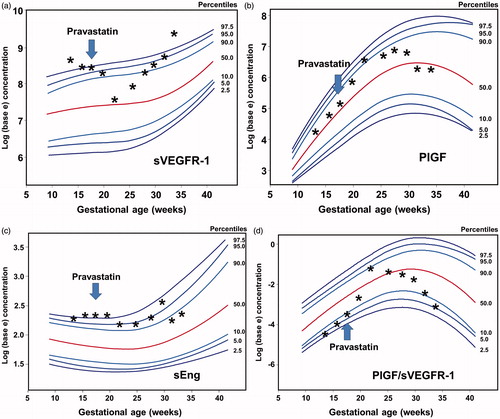
Table 1. Plasma concentrations (percentile for gestational age) of angiogenic and anti-angiogenic factors.
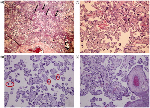
Figure 3. Histopathological examination of the placenta from the current pregnancy reveals fibrinoid deposition (arrow) in the intervillous space, involving 20% of the villi (a) and areas with normal intervillous space (b) (H&E, 100×). Small and poorly developed villi (circle) or distal villous hypoplasia (H&E, 100×) are shown in (c). (d) Displays well-developed villi at 34 weeks of gestation (H&E, 100×).