Figures & data
Figure 1. Custom-made ankle foot orthosis designed to measure dorsi and plantar flexors muscle force.
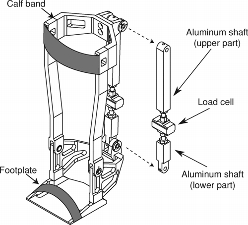
Table 1. Subject characteristics.
Figure 2. Baseline strength of i) dorsiflexors, ii) plantar flexors and iii) quadriceps for maximal voluntary contraction (MVC –panel A) and potentiated twitch force (Twpot –panel B) in patients with COPD (filled bars) and healthy control subjects (open bars). Values are mean ± SD. * p < 0.05 vs. controls.
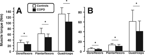
Table 2. End-exercise cardiorespiratory response to standardized endurance walking test.
Figure 3. Post-exercise loss in strength for the i) dorsiflexors, ii) plantar flexors and iii) quadriceps for maximal voluntary contraction (MVC –panel A) and potentiated twitch force (Twpot –panel B) in patients with COPD (filled bars) and healthy control subjects (open bars). Values are mean ± SD and expressed as the% fall from resting values. * p < 0.05 vs. controls.
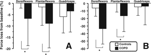
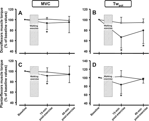
Figure 4. Post-exercise loss in strength for the i) dorsiflexors and ii) plantar flexors for maximal voluntary contraction (MVC –panel A and C) and potentiated twitch force (Twpot –panel B and D) in patients with COPD (filled circles) and healthy control subjects (open circles) at 15 minutes and 40 minutes post-exercise. Values are mean ± SD. The values at 40 minutes post-exercise are only available for 8 patients with COPD (n = 8) and 6 healthy controls (n = 6). * p < 0.05 vs. baseline force values within groups.