Figures & data
Figure 1. Establishment of hUCB CD34+ cell co-cultures with AFT024 (a-c), proliferation of progenitor cells (d) and phenotypic behavior along the 4 week period (e). a) CD34+ cells from hUCB after MACS enrichment; b) monolayer of stroma cells after irradiation (day 0); c) CD34+ enriched cells over stroma AFT024 (day 0); d) Cobblestone-like areas over feeder layer (day 7); e) percentage of CD34+ cells obtained by flow cytometry after each week of culture; p < 0.001.
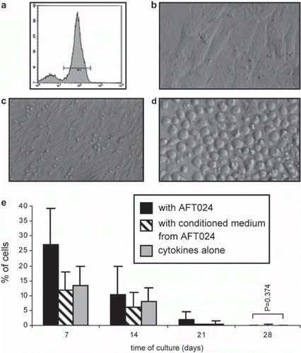
Table 1. Percentage of CD56+ CD3− and proportion of CD56+ CD3−KIR+ , CD56+ CD3−NCR+ and CD56+ CD3−NKG2A/CD94− cells generated by CD34+ cells grown in the described three conditions. Kinetics of expression along the time can be better visualized in .
Figure 2. Phenotype of in vitro cultured hUCB CD34+ cells. Flow cytometry analysis after 7 (a) and 28 (b) days of culture. Note that the CD34+ cells almost become extinct between day 7 (d7) and day 28 (d28) with the simultaneous appearance of CD56+ phenotype. Differentiation areas can be seen in c) at day 14 (d14) of culture. Detailed data is shown in .
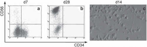
Figure 3. Flow cytometry analysis of differentiated hUCB CD34+ cells at the end of the experimental period. Cells were analysed by flow cytometry and positivity for the different mAbs was defined in the morphological gate represented in a). NK cells population is shown in b) and the expression several receptors are shown in c) for the KIRmix, in d) for the NCRmix and in e) for NKG2A.
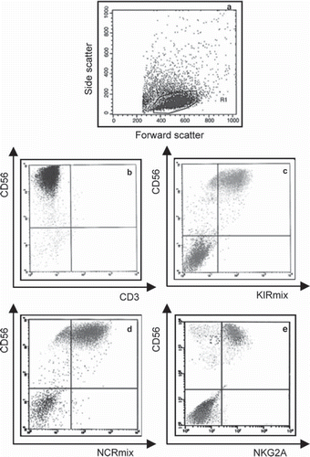
Figure 4. hUCB CD34+ cells differentiation into NK cells along the 4-week period. Cells were analysed by flow cytometry and positivity for the different mAbs was defined in the morphological lymphoid gate defined in . Results from a) and b) were additionally defined in the CD56+ CD3+ gated region (see ) and results from d) were also defined in the CD56+ CD94+ gated region (data not showed). The graphics show the CD56+ CD3+ cells generated in vitro from CD34+ progenitors (a) and the kinetics of expression of KIRs (b), NKps (c) and CD94/NKG2A- (d) (p < 0.001). See for detailed data.
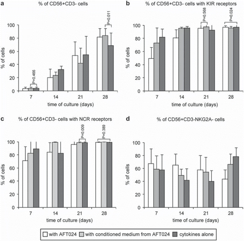
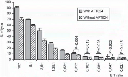
Figure 5. Cytotoxicities mediated by in vitro differentiated NK cells derived from hUCB-HSCs (effector cells) against K-562 target cells. These activities were measured using a Non-Radioactive Cytotoxicity test at the effector:target (E:T) cell ratios indicated. Each data bar represents a mean SD percentage value of five separate experiments, each performed in quadruplicate (p < 0.05).