Figures & data
Figure 1. Schematic representation of the molecular constituents of the desmosome and their significance in pemphigus autoimmunity. The first reports describing desmosomal molecules as target antigens of pemphigus have been listed in the figure. For example, Stanley et al. reported in 1986 that Dsg1 was targeted by pemphigus autoimmunity, i.e. patients with pemphigus developed anti-Dsg1 IgG. Note that later studies have confirmed and further investigated the role of these antigens in the pathogenesis of pemphigus. Dsc, desmocollin; Dsg, desmoglein; DP, desmoplakin; Pg, plakoglobin; Pkp, plakophilin.
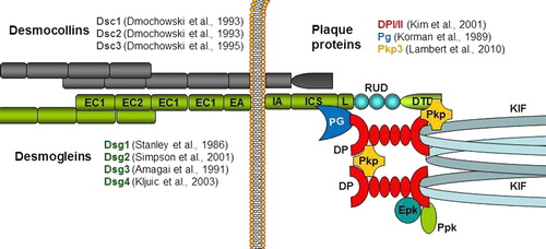
Figure 2. Intracellular pathways involved in the disruption of desmosomal adhesion in pemphigus. The acantholytic mechanisms target adhesion molecules as a result of both direct processing and indirect intracellular signaling. They include the internalization and degradation of Dsg3, apoptotic signals that lead to activation of caspases (e.g., Casp3) and metalloproteinases (e.g., MMP9), hyper-adhesion, and transcriptional/translational down-regulation of desmosomal molecules. As shown in the picture, most of the changes are orchestrated by protein kinases. Dsgs, desmogleins; Dscs, desmocollins; IFs, intermediate filaments; Casp3, Caspase 3; MMP9, matrix metalloproteinase 9.
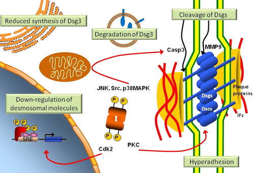
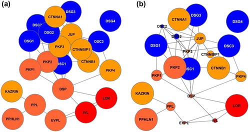
Figure 3. Simplified desmosomal interactome in pemphigus. (a) Graphical representation of the intrinsic components of the desmo-adhesome (with the exception of keratins). The intrinsic desmosomal proteins are depicted as circles (nodes) that are connected by lines (interactions) based on their reciprocal binding interactions, thus forming a network. Nodes have been assigned different colors according to the function of the respective proteins: transmembrane (blue), adaptors (orange), and cytoskeleton molecules (red); (b) the relative gene expression changes of the same molecules after perturbation of the system in pemphigus vulgaris (PV) have been represented as differential size of the nodes (e.g., down-regulated molecules have proportionally smaller size). The original microarray data are from a mouse model of PV (CitationLanza et al., 2008).