Figures & data
Figure 1. IFNγ production by hTERT-reactive T-lymphocytes. Matured dendritic cells were loaded with nonapeptide antigen (139TSAPDTRPA147) derived from hTERT protein. Autologous T-lymphocytes were stimulated with those antigen-pulsed DCs. Seven days after the initial stimulation, T-lymphocytes were re-stimulated with the same antigen-pulsed DCs. HIV-1-derived peptide (476ILKEPVHGV484) was used as a negative control. Activation of (A) CD3+CD4+ and (B) CD3+CD8+ T-lymphocytes in an experimental (hTERT) and control (HIV-1) group was measured by IFNγ production. Flow cytometry analysis was performed after 24 h of re-stimulation with hTERT- or HIV-1-pulsed DCs. Data are reported as mean (±SD). Significant differences between groups (at p < 0.01) are denoted with an asterisk.
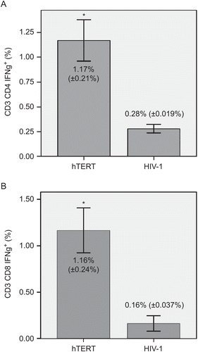
Figure 2. Separation of hTERT-reactive T-lymphocytes. Comparison of IFNγ+ subsets of (A) CD3+CD4+ and (B) CD3+CD8+ T-lymphocytes in positive and negative fractions. Immunomagnetic separation was performed twice. Panels show percentages of IFNγ-producing cells of each subset. Data are shown as mean (±SD). Significant differences between groups (at p < 0.01) are denoted with an asterisk.
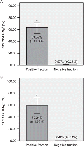
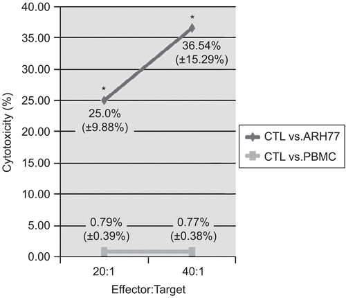
Figure 3. Cytotoxic activity of hTERT-specific CTLs against the ARH77 myeloma cell line. T-lymphocyte-mediated cytotoxicity was measured using the expanded hTERT-reactive T-lymphocytes as effector (E) cells. DiOC-labeled hTERT-positive ARH77 myeloma cells were used as the target cells (T). To validate the specificity of the effector cells, PBMCs were used as a negative control target. Data are shown as mean (±SD) cytotoxic activities at the E:T ratios shown. Significant (at p < 0.05) differences between results with the different target cell types are denoted with an asterisk.