Figures & data
Figure 1. Effects of TBBPA exposures on the ability of NK cells to lyse or bind tumor cells. NK cells were exposed to 0.05–5 μM TBBPA for (A) 24 h, (B) 48 h, or (C) 6 days, and the tested for lytic function (closed diamonds) or binding function (closed squares). To combine results from separate experiments (using cells from different donors), levels of lysis or binding were normalized as the percentage of the lytic or binding function of the control cells (untreated cells from the same donor) in a given experiment. Results were pooled from three separate experiments using different donors (triplicate determinations for each experiment; n = 9; mean ± SD). Significant decreases in Lytic Function as compared to control: (A) 0.5, 1, 2.5, and 5 μM (P < 0.001); (B) 0.5, 1, and 2.5 μM (P < 0.0001); and (C) 0.1, 0.25, 0.5, and 1 μM at (P < 0.01). Significant decreases in Binding Function as compared to control: (A) 2.5 and 5 μM (P < 0.0001); (B) 1 and 2.5 μM (P < 0.05); and (C) 1 μM (P < 0.01).
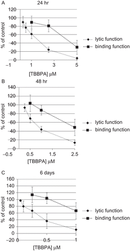
Figure 2. Time course of the effects of 0.5 μM (black bar) and 1 μM (gray bar) TBBPA on the ability of NK cells to lyse tumor cells. Results are the 0.5 and 1 μM data from plotted vs. length of exposure.
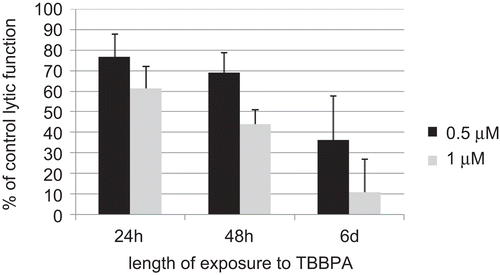
Figure 3. Effects of a 1-h exposure to TBBPA followed by various periods in TBBPA-free media on the ability of NK cells to lyse or bind tumor cells. NK cells were exposed to 0.5–10 μM TBBPA for 1 h, and then tested for lytic function (closed diamonds) or binding function (closed squares) after (A) 24 h, (B) 48 h, or (C) 6 days. Results were normalized as described in . Significant decreases in Lytic Function as compared to control: (A) 1, 2.5, 5, and 10 μM (P < 0.001); (B) 2.5, 5, and 10 μM (P < 0.05); (C) 2.5, 5, and 10 μM at (P < 0.01). Significant decreases in Binding Function as compared to control: (A) none; (B) none; and (C) 10 μM (P < 0.0001).
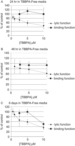
Figure 4. Time course of the effects of 1-h exposures to 5 μM (black bar) and 10 μM (gray bar) TBBPA followed by a period in TBBPA-free media on the ability of NK cells to lyse tumor cells. Results are the 5 and 10 μM data from plotted vs. length of time in TBBPA-free media.
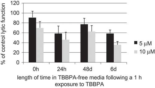
Figure 5. Effects of TBBPA exposures on the ATP levels and lytic function of NK cells. NK cells were exposed to 0.05–10 μM TBBPA for (A) 24 h, (B) 48 h, or (C) 6 days, and then tested for ATP levels (closed squares) or lytic function (closed diamonds). Results were normalized as described in . Significant decreases in ATP levels as compared to control: (A) 5 μM (p < 0.0001); (B) 1 and 2.5 μM (P < 0.0001); and (C) 0.25, 0.5, and 1 μM (P < 0.001). Significant decreases in Lytic Function as compared to control: (A) 0.5, 1, 2.5, and 5 μM (P < 0.001); (B) 0.5, 1, and 2.5 μM (P < 0.0001); and (C) 0.1, 0.25, 0.5, and 1 μM at (P < 0.01).
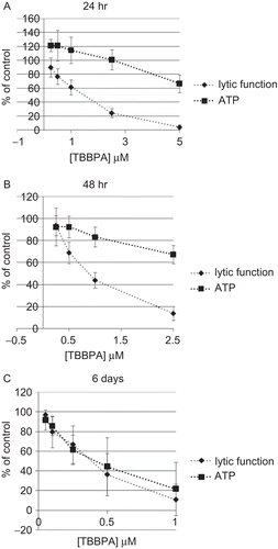
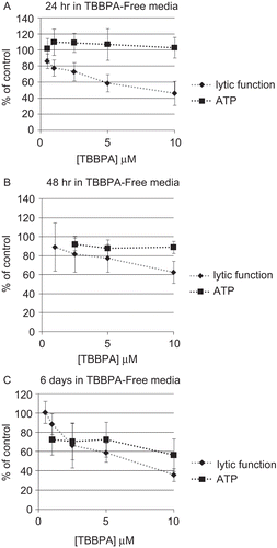
Figure 6. Effects of TBBPA exposures on the ATP levels and lytic function of NK cells. NK cells were exposed to 0.5–10 μM TBBPA for 1 h, followed by periods of time in TBBPA-free media before assaying for ATP levels (closed squares) or lytic function (closed diamonds). (A) 24 h, (B) 48 h, or (C) 6 days. Results were normalized as described in . Significant decreases in ATP Levels as compared to control: (A) none; (B) 2.5, 5, and 10 μM (P < 0.05); and (C) 1, 2.5, 5, and 10 μM (P < 0.01). Significant decreases in Lytic Function as compared to control: (A) 1, 2.5, 5, and 10 μM (P < 0.001); (B) 2.5, 5, and 10 μM (P < 0.05); (C) 2.5, 5, and 10 μM (P < 0.01).