Figures & data
Figure 1. Cell viability of lymphocytes following 24 (upper) and 48 (lower) h of exposure to varying concentrations of Cr6+ and Co2+ ions, as measured by neutral red (NR) assay. (A and C) Resting and (B and D) anti-CD3-activated lymphocytes. A value of 100% indicates unexposed control cells; results are means ± SE (n = 18). *Significantly different from control values (at p < 0.05) by one-way analysis of variance (ANOVA) followed by Dunnett’s multiple comparison test. †Significantly different from Co2+ values (at p < 0.05) by two-sample t-test.
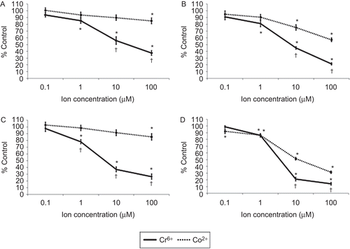
Figure 2. T-lymphocyte proliferation following anti-CD3 ± anti-CD28 activation and exposure to varying concentrations of Cr6+ and Co2+ ions for 48 h. A value of 100% indicates unexposed control cells; results are means ± SE (n = 12). *Significantly different from control values (at p < 0.05) by one-way analysis of variance (ANOVA) followed by Dunnett’s multiple comparison test.
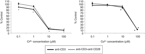
Figure 3. IL-2 release from anti-CD3 ± anti-CD28 activated lymphocytes exposed to varying concentrations of Cr6+ and Co2+ ions for 48 h. Results are means ± SE (n = 3). *Significantly different from control values (at p < 0.05) by one-way analysis of variance (ANOVA) followed by Dunnett’s multiple comparison test.
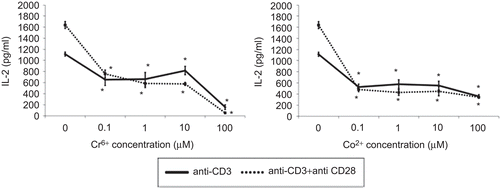
Figure 4. IFNγ release from anti-CD3 ± anti-CD28 activated lymphocytes exposed to varying concentrations of Cr6+ and Co2+ ions for 48 h. Results are means ± SE (n = 3). *Significantly different from control values (at p < 0.05) by one-way analysis of variance (ANOVA) followed by Dunnett’s multiple comparison test.
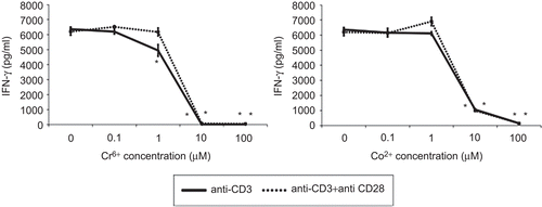
Figure 5. Resting and anti-CD3 activated lymphocytes stained with annexin V and 7-AAD following exposure to 100 µM Cr6+ and 100 µM Co2+ for 24 h. Viable cells were annexin V− and 7-AAD−. Cells in early stages of apoptosis were annexin V+ but 7-AAD−, whereas cells in late stages of apoptosis were both annexin V+ and 7-AAD+.
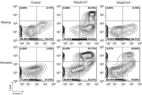
Figure 6. Total apoptosis in lymphocytes following 24 (upper) and 48 (lower) h of exposure to Cr6+ and Co2+. (A and C) Resting and (B and D) anti-CD3-activated lymphocytes. Results are mean (± SE; n = 6) proportion of lymphocytes expressing annexin V. A value of 100% indicates baseline apoptosis in unexposed control cells. *Significantly different from control values (at p < 0.05) by one-way analysis of variance (ANOVA) followed by Dunnett’s multiple comparison test. †Significantly different from Co2+ values (at p < 0.05) by two-sample t-test.
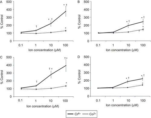
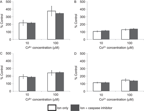
Figure 7. Total apoptosis in lymphocytes (pre-treated for 24 h with/without caspase-3 inhibitor (50 µM Z.Devd.FMK)) following 24 h of exposure to Cr6+ or Co2+. (A and C) Resting and (B and D) anti-CD3-activated lymphocytes. Results are mean (± SE; n ≥ 3) proportion of lymphocytes expressing annexin V. A value of 100% indicates baseline apoptosis in unexposed control cells.