Figures & data
Figure 1. Chemical structure of triclosan.
Figure 2. Allergic sensitization potential after dermal exposure to triclosan. Analysis of allergic sensitization potential of triclosan using LLNA. Disintegration per minute (DPM) represent [3H]-thymidine incorporation into draining lymph node cells of BALB/c mice following exposure to vehicle or concentration of triclosan (0.75–3.0%). SI value is stimulation index (fold-change over vehicle control). Bars shown are means (±SE) of five mice per group.
![Figure 2. Allergic sensitization potential after dermal exposure to triclosan. Analysis of allergic sensitization potential of triclosan using LLNA. Disintegration per minute (DPM) represent [3H]-thymidine incorporation into draining lymph node cells of BALB/c mice following exposure to vehicle or concentration of triclosan (0.75–3.0%). SI value is stimulation index (fold-change over vehicle control). Bars shown are means (±SE) of five mice per group.](/cms/asset/f9b560e8-54f7-4c35-b144-a387020652d7/iimt_a_1029146_f0002_b.jpg)
Table 1. Body\organ weights of female B6C3F1 mice dermally exposed to triclosan for 28 days.
Table 2. Hematology parameters of female B6C3F1 mice dermally exposed to triclosan for 28 days.
Figure 3. Effects of dermal exposure to triclosan on lymph node and spleen cellularity. Effects of dermal exposure to triclosan for 28 days on (A) lymph node and (B) spleen cellularity in female B6C3F1 mice. Values shown are means (±SE) for each group. Levels of statistical significance are denoted *p < 0.05 and **p < 0.01 as compared to acetone vehicle.
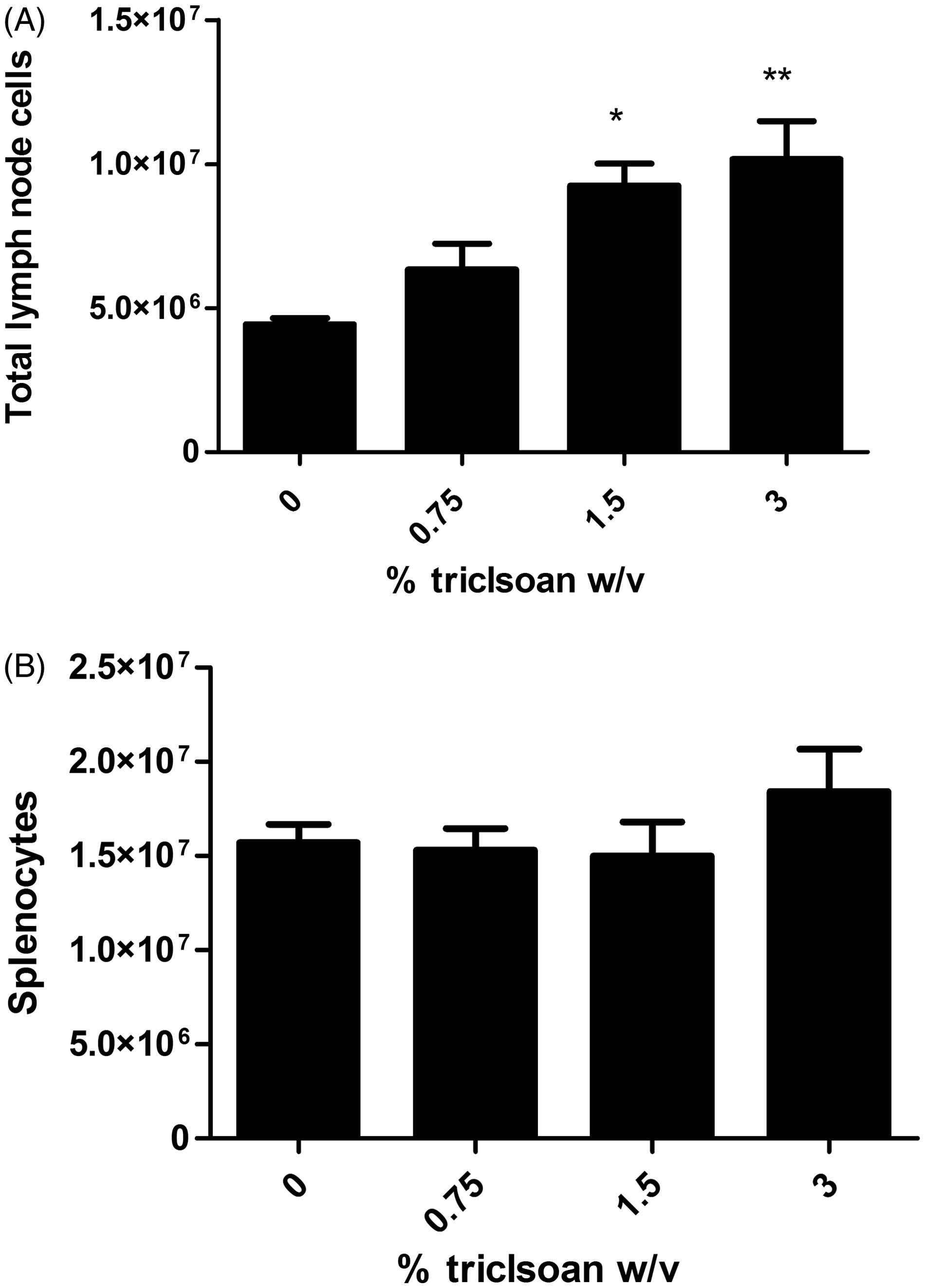
Figure 4. Effects of dermal exposure to triclosan for 28 days on leukocyte populations. Effects of dermal exposure to triclosan for 28 days on (A) total cell number and (B) frequency of lymphocyte subpopulations in female B6C3F1 mice. Numbers of B-cells, CD4+ and CD8+ T-cells, dendritic cells and NK cells were enumerated using flow cytometry. Values shown are means (±SE) for each group. Levels of statistical significance are denoted *p < 0.05 and **p < 0.01 as compared to acetone vehicle.
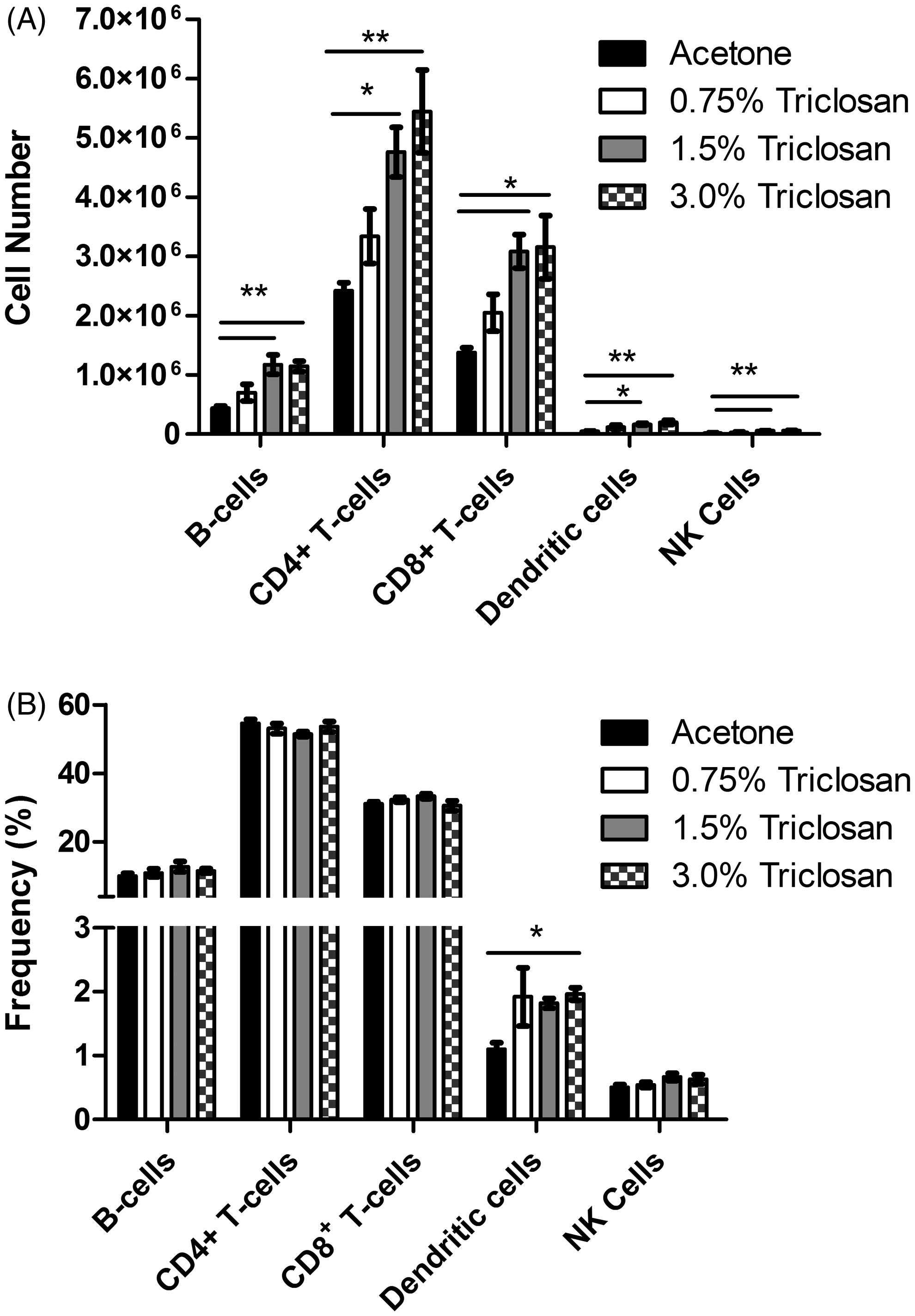
Figure 5. Effects of dermal exposure to triclosan for 28 days on splenocyte populations. Effects of dermal exposure to triclosan for 28 days on (A) total cell number and (B) frequency of lymphocyte subpopulations in female B6C3F1 mice. Numbers of B-cells, CD4+ and CD8+ T-cells, dendritic cells and NK cells were enumerated using flow cytometry. Values shown are means (±SE) for each group.
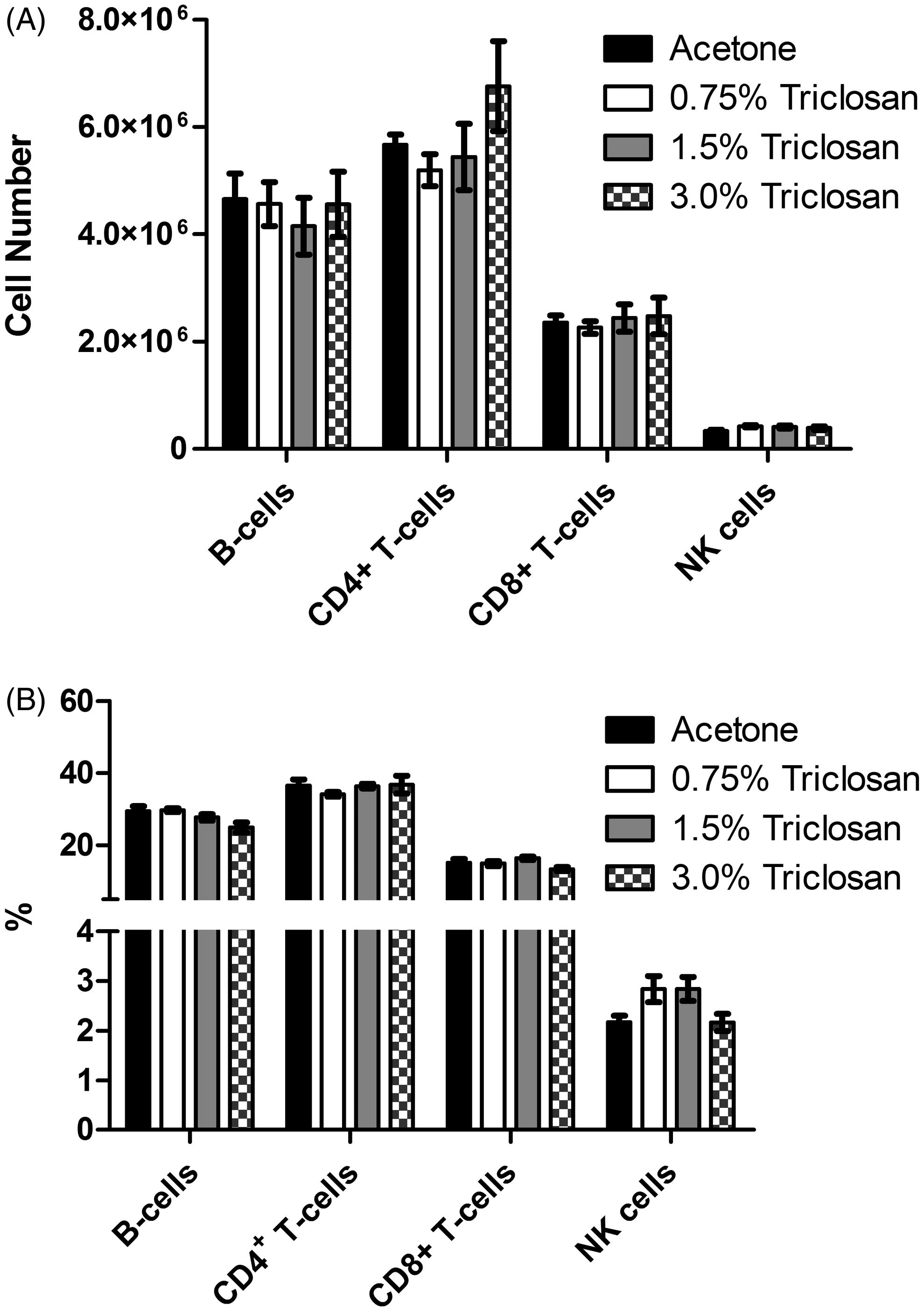
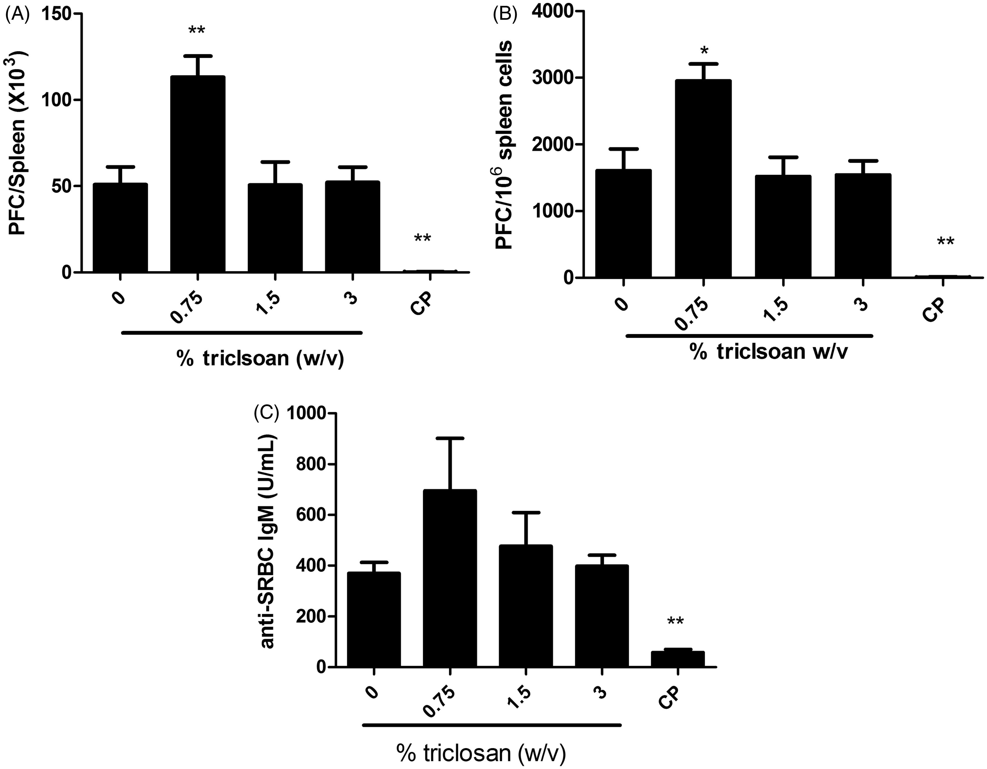
Figure 6. Triclosan does not suppress the spleen IgM response to SRBC. Analysis of antibody producing cells after a 28-day dermal exposure to triclosan on the (A) total and (B) specific activity IgM response to SRBC in the spleen and serum. (C) Bars represent mean fold-change (±SE) of six mice/group. Cyclophosphamide (CP) was included as the positive control. Levels of statistical significance are denoted *p < 0.05 and **p < 0.01 as compared to acetone vehicle.