
Figures & data
Table 1. Demographic and clinical variables in both AIH and PSC groups.
Table 2. Histopathology variables in both AIH and PSC groups.
Figure 1. Composite photomicrograph. A&B were taken from a patient with autoimmune hepatitis and C&D from another patient with primary sclerosing cholangitis. A. H&E-stained slide with a 4× lens demonstrates a cirrhotic liver with bands of bridging fibrosis and regenerative nodules. B. Trichrome-stained liver with a 4× lens taken at the same area as the H&E-stained section from A highlighting the bridging fibrosis in blue. C. H&E-stained slide with a 40× lens showing extensive chronic inflammation extending beyond the limiting plate and a few compressed bile ducts. D. H&E-stained slide with a 40× lens depicts moderate chronic inflammation surrounding the bile duct.
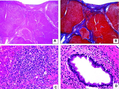
Table 3. Clinical laboratory variables in both AIH and PSC groups.
Table 4. Autoimmunity laboratory markers in both AIH and PSC groups.