Figures & data
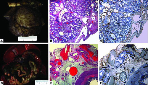
Figure 1. Composite photograph with gross and photomicrographs from the autopsy. A. The lungs after 24 h of formalin fixation. When fresh, they were pale pink with a slightly bumpy pleural surface. The aorta is horizontally placed at the upper portion of the photograph. B. Hematoxylin-and eosin (H&E)-stained photograph taken from the lung with the 4× -lens depicts the pleural surface and part of the pulmonary parenchyma. Notice the dilated vessels. C. Same field of view and magnification as in 1-B, but stained by immunohistochemistry for D2-40 highlights the lymphatic origin of dilated vessels. D. The upper part of the abdominal organs demonstrates the liver with gallbladder, intestine and congested mesenteric vessels. E. The H&E-stained photomicrograph from intestine and mesentery with the 4× -lens shows the intestine in the right lower corner and numerous dilated mesenteric vessels. F. Same field of view and magnification as in 1-E, but stained by immunohistochemistry for D2-40 highlights the lymphatic nature of dilated vessels.