1,730
Views
33
CrossRef citations to date
0
Altmetric
Original Articles

Experimental 70% hydrofluoric acid burns: histological observations in an established human skin explants ex vivo model

, , , , , , & show all
Pages 100-107 | Received 21 Aug 2010, Accepted 15 Oct 2010, Published online: 15 Nov 2010

Figures & data

Figure 1.  Chemical reactions between fluoride ions and calcium/magnesium.

Figure 1.  Chemical reactions between fluoride ions and calcium/magnesium.

Table 1.  Mean thickness of the human skin explants layers.

Figure 2.  Normal aspect of skin optical microscopy with 40× objective.

Figure 2.  Normal aspect of skin optical microscopy with 40× objective.

Figure 3.  Skin histological aspect after a 20-sec exposure to 30 µL of 70% hydrofluoric acid (HF). No deterioration of the epidermal or dermal structures.
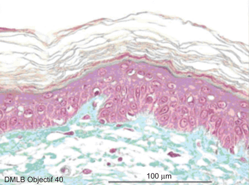
Figure 3.  Skin histological aspect after a 20-sec exposure to 30 µL of 70% hydrofluoric acid (HF). No deterioration of the epidermal or dermal structures.

Figure 4.  Skin histological aspect at 1 min after a 20-sec exposure to 30 µL of 70% hydrofluoric acid (HF). The epidermis showed four to five cellular layers with a slightly modified morphology. The cells showed gray cytoplasm in the upper layer and the nuclei became pyknotic. The cellular structures of the basal epidermis and throughout the dermis showed normal morphologies.
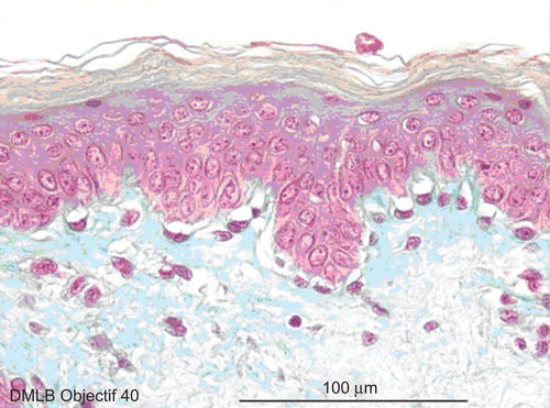
Figure 4.  Skin histological aspect at 1 min after a 20-sec exposure to 30 µL of 70% hydrofluoric acid (HF). The epidermis showed four to five cellular layers with a slightly modified morphology. The cells showed gray cytoplasm in the upper layer and the nuclei became pyknotic. The cellular structures of the basal epidermis and throughout the dermis showed normal morphologies.

Figure 5.  Skin histological aspect at 2 min after a 20-sec exposure to 30 µL of 70% hydrofluoric acid (HF). The skin showed four to five cellular layers with definitely abnormal morphology: cells with nuclei becoming pyknotic, especially in the higher epidermal layers, and the cytoplasm becoming acidophilic as reflected by orange keratinocyte pigmentation. The cellular structures in the dermis showed normal morphology.
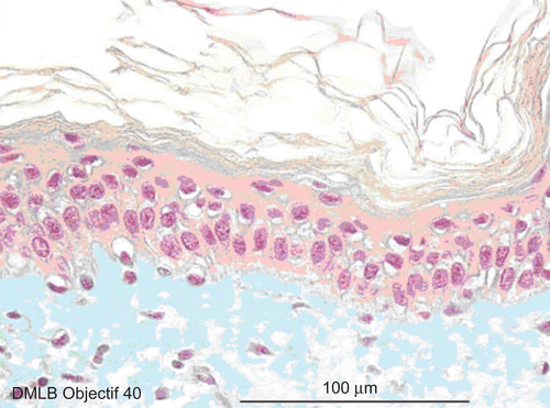
Figure 5.  Skin histological aspect at 2 min after a 20-sec exposure to 30 µL of 70% hydrofluoric acid (HF). The skin showed four to five cellular layers with definitely abnormal morphology: cells with nuclei becoming pyknotic, especially in the higher epidermal layers, and the cytoplasm becoming acidophilic as reflected by orange keratinocyte pigmentation. The cellular structures in the dermis showed normal morphology.

Figure 6.  Skin histological aspect at 3 min after a 20-sec exposure to 30 µL of 70% hydrofluoric acid (HF). Lesions of four to five epidermis cellular layers were characterized by numerous cells with moderately pyknotic nuclei and edema surrounding the nuclei. At the base of the stratum corneum and in the basal epidermis layer, cells showed characteristic cytoplasmic alterations. In the papillary dermis, the cellular structures were slightly pyknotic. The reticular dermis remained normal.
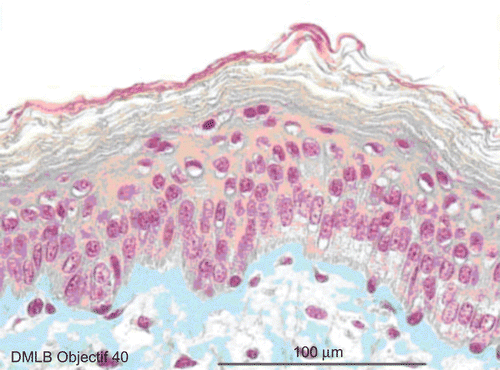
Figure 6.  Skin histological aspect at 3 min after a 20-sec exposure to 30 µL of 70% hydrofluoric acid (HF). Lesions of four to five epidermis cellular layers were characterized by numerous cells with moderately pyknotic nuclei and edema surrounding the nuclei. At the base of the stratum corneum and in the basal epidermis layer, cells showed characteristic cytoplasmic alterations. In the papillary dermis, the cellular structures were slightly pyknotic. The reticular dermis remained normal.

Figure 7.  Skin histological aspect at 4 min after a 20-sec exposure to 30 µL of 70% hydrofluoric acid (HF). The same deteriorations in the epidermis were observed compared with those described at 3 min. In the papillary dermis, cells more clearly showed pyknotic nuclei, but the reticular dermis remained normal.
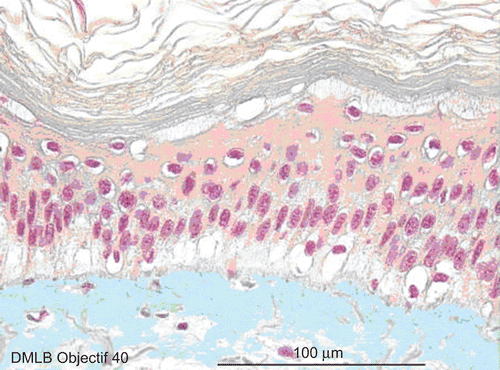
Figure 7.  Skin histological aspect at 4 min after a 20-sec exposure to 30 µL of 70% hydrofluoric acid (HF). The same deteriorations in the epidermis were observed compared with those described at 3 min. In the papillary dermis, cells more clearly showed pyknotic nuclei, but the reticular dermis remained normal.

Figure 8.  Skin histological aspect at 5 min after a 20-sec exposure to 30 µL of 70% hydrofluoric acid (HF). The same lesions as those detected after 4 min were observed in the epidermis and papillary dermis. However, as the HF had penetrated into the reticular dermis, slightly pyknotic nuclei were observed in cells in this deepest layer.
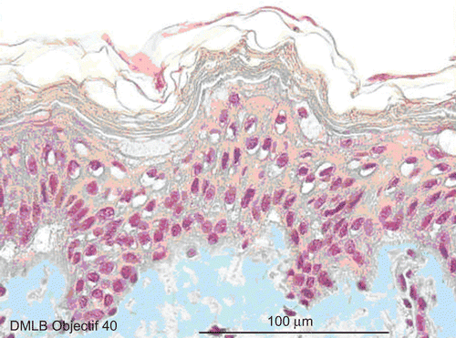
Figure 8.  Skin histological aspect at 5 min after a 20-sec exposure to 30 µL of 70% hydrofluoric acid (HF). The same lesions as those detected after 4 min were observed in the epidermis and papillary dermis. However, as the HF had penetrated into the reticular dermis, slightly pyknotic nuclei were observed in cells in this deepest layer.

Table 2.  Comparison between known values of skin thickness.

Table 3.  Comparison of thickness of abdomen skin between races.

Table 4.  Dynamics of appearance of lesions after skin exposure to 70% hydrofluoric acid (HF).

Table 5.  Details of histological cell alterations during a 5-min skin exposure to 70% hydrofluoric acid (HF).
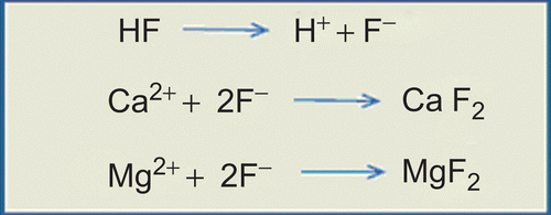
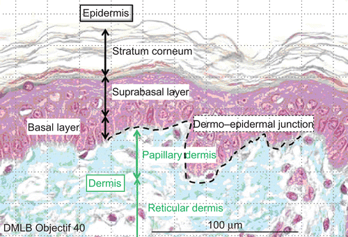
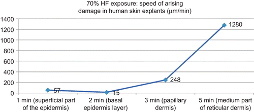
Figure 9.  Kinetics of 70% hydrofluoric acid (HF) through human ex vivo skin explants.

Figure 9.  Kinetics of 70% hydrofluoric acid (HF) through human ex vivo skin explants.