Figures & data
Table 1. Pain and skin lesions: depending on HF concentration.
Table 2. Explants and treatment groups.
Figure 1. (A) and (B) Exposed 70% HF, nonwashed explants, aspect at 24 h after a 20-sec exposure. Epidermis (A) and dermis (B). At 24 h after a 20-sec exposure, the epidermis presented completely necrotic structures with the appearance of coagulation necrosis. The lesions were less intense in the papillary and reticular dermis
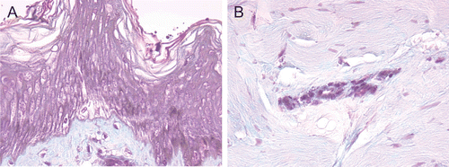
Figure 2. (A) and (B) HF-exposed explants rinsed with water + CaG, aspect at 24 h after a 20-sec exposure. Epidermis (A) and dermis (B). Edematous changes were visible in the epidermal basal layer at 24 h after a 20-sec exposure. An appearance of coagulation necrosis including pyknotic nuclei and acidophilic cytoplasm was noted in the papillary dermis. These changes were less apparent in the reticular dermis.
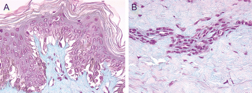
Figure 3. (A) and (B) HF-exposed explants rinsed with Hexafluorine®; aspect at 24 h after a 20-sec exposure. Epidermis (A) and dermis (B). No deterioration of the structures of either the epidermis or dermis was observed.
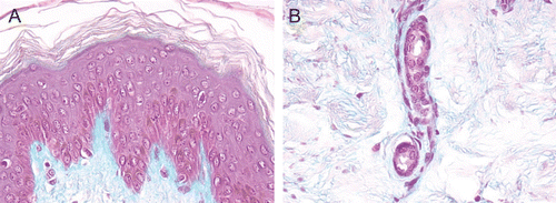
Table 3. Schematic summary of the histological results for all the groups and for each skin layer.