939
Views
25
CrossRef citations to date
0
Altmetric
Research Article

Treatment and outcome of giant cell tumors of the pelvis

20 cases followed for 1 to 11 years

, , , , &
Pages 590-596 | Accepted 12 May 2009, Published online: 16 Nov 2009

Figures & data

Data on 20 patients with giant cell tumor of bone

Figure 1. Localization of main tumor components. 3 groups according to Enneking and Dunham (Citation1978). Region I: iliosacral area; region II: acetabular area; region III: ischiopubic area. 9 tumors affected region I, 6 affected region II, and 5 affected region III.
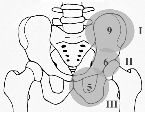
Figure 1. Localization of main tumor components. 3 groups according to Enneking and Dunham (Citation1978). Region I: iliosacral area; region II: acetabular area; region III: ischiopubic area. 9 tumors affected region I, 6 affected region II, and 5 affected region III.

Figure 2. Case 2. a. GCT of the left ilium with sacral extension and proximal cortical breakthrough. b. After intralesional curettage and bone cement packing of the defect.
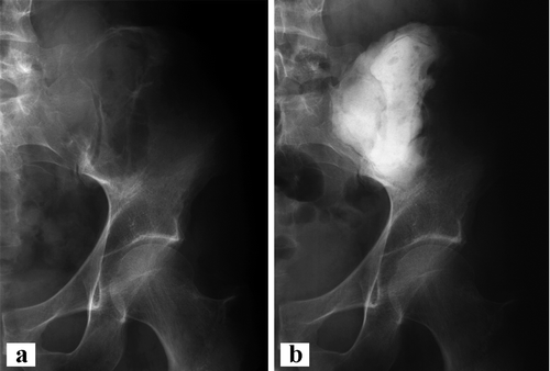
Figure 2. Case 2. a. GCT of the left ilium with sacral extension and proximal cortical breakthrough. b. After intralesional curettage and bone cement packing of the defect.

Figure 3. Case 20. a. GCT of the right pubic bone with bony destruction. b. After intralesional resection of pubic bone, including the symphysis. Proximal margins were additionally curetted and cemented. Note the multiple hemostatic clips.
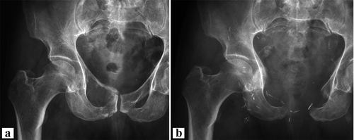
Figure 3. Case 20. a. GCT of the right pubic bone with bony destruction. b. After intralesional resection of pubic bone, including the symphysis. Proximal margins were additionally curetted and cemented. Note the multiple hemostatic clips.

Figure 4. Case 7. a. Giant cell tumor of the left iliac wing. b. After wide resection of the left iliac wing without reconstruction.
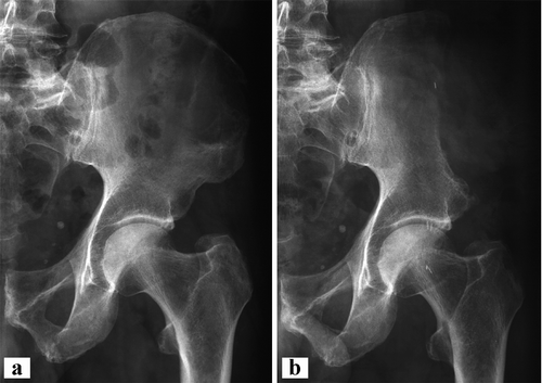
Figure 4. Case 7. a. Giant cell tumor of the left iliac wing. b. After wide resection of the left iliac wing without reconstruction.

Figure 5. Case 11. a. After intralesional resection of a GCT of the ischium. Additional curettage and bone cement packing of the acetabular extension was performed. The cement was fixed with 2 screws to prevent dislocation. b. CT showing dislocation of the dorsal screw, necessitating surgical removal.
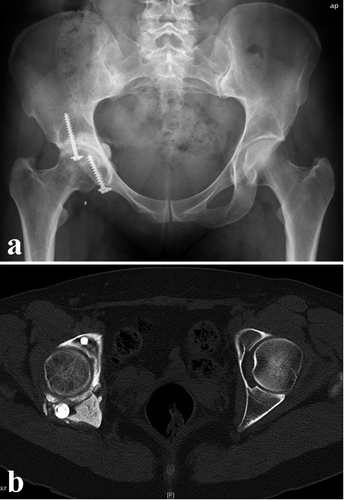
Figure 5. Case 11. a. After intralesional resection of a GCT of the ischium. Additional curettage and bone cement packing of the acetabular extension was performed. The cement was fixed with 2 screws to prevent dislocation. b. CT showing dislocation of the dorsal screw, necessitating surgical removal.