Figures & data
Details of revised hip resurfacings including patient demographics and duration “in situ” for each group of patients
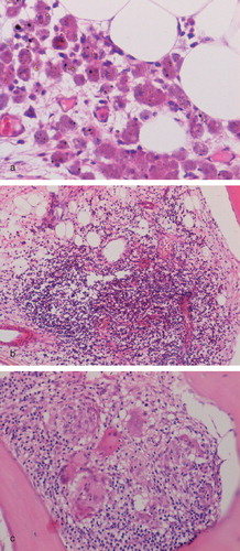
Figure 1. Necrosis in peri-implant soft tissues of an MoM hip resurfacing. (a) Necrosis of superficial fibrous tissue that is covered by fibrin in a case of component loosening. (b) Coagulative necrosis in which ghost-like outlines of plump macrophages are evident in a case of pseudotumor.
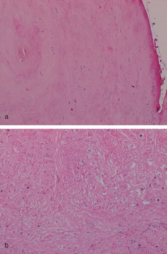
Figure 2. Inflammation in the pseudomembrane from a case of component loosening showing (a) macrophage infiltration and metal particles (arrowed); (b) macrophage and giant cell expression of CD68; and (c) discrete granuloma formation.
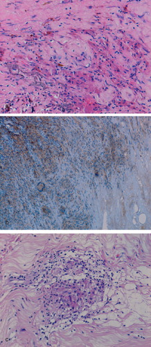
Figure 3. Macrophage and giant cell (arrowed) granulomatous response around an area of coagulative necrosis (top) in a case of MoM-implant associated pseudotumor.
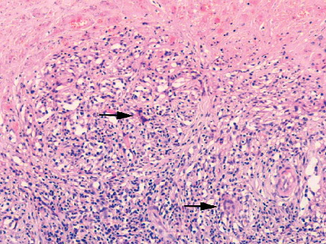
Figure 4. Pseudomembrane from a case of MoM implant component loosening showing (a) numerous perivascular lymphoid aggregates and a diffuse lymphocyte infiltrate; (b) expression of CD3 by lymphocytes in the diffuse inflammatory cell infiltrate; and (c) CD20-expressing B lymphocytes in lymphoid aggregates.
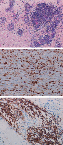
Figure 5. Femoral bone marrow from a revised MoM hip resurfacing showing (a) a collection of macrophages containing metal particles (arrowed); (b) a small lymphoid aggregate; and (c) a discrete macrophage and giant cell granuloma.