Figures & data
Figure 1. Histological features of osteonecrosis in rabbits. A. Nomal bone harvested from a comparable rabbit not treated with α-tocopherol nor treated with methylprednisolone acetate. B. Typical osteonecrotic lesion (experimental group). C. Nomal bone marrow cells. D. Necrotic bone marrow cells. Bone marrow cells had necrosis and stained acidophilic. The nuclei of bone marrow cells displayed pyknosis and karyorrhexis. The cellular structure of fat cells collapsed. E. Normal bone. Empty lacunae of the osteocytes (arrowhead) without bone marrow cell necrosis or fat cell necrosis. F. Osteonecrotic bone. Bone cells in the bone trabeculae showed pyknosis and empty lacunae (arrow) that were associated with necrotic changes of the surrounding bone marrow cells. Stain: hematoxylin and eosin; magnification: ×200 (A, B), ×400 (C, D, E, F).
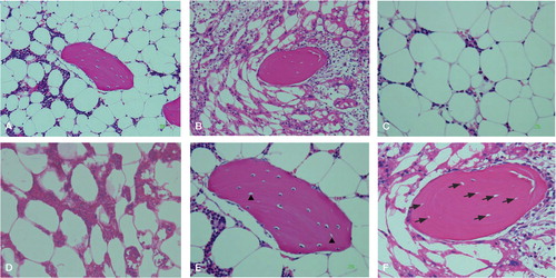
Figure 2. A. Strong immunoreactivity found in both a marrow vessel and bone marrow cells. B. Weak immunoreactivity found in both a marrow vessel and bone marrow cells. Stain: anti-malondialdehyde monoclonal antibody (clone 1F83); magnification: ×200.
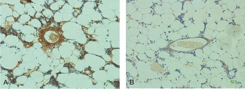
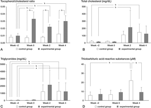
Figure 3. Hematological findings. The tocopherol/cholesterol ratios (A), total cholesterol levels (B), triglyceride levels (C), and concentration of thiobarbituric acid-reactive substances (D) are shown. a P < 0.05, b P < 0.01 by Tukey-Kramer method for between-group and within-group comparisons).