Figures & data
Figure 1. Schematic representation of the muscle damage measurements relative to the midsubstance cross-sectional surface area (MCSA). A relatively large damaged area at the origo (α1) may yield the same percentage of damaged MCSA (α) as a relatively small area at the musculotendinous insertion (α2).
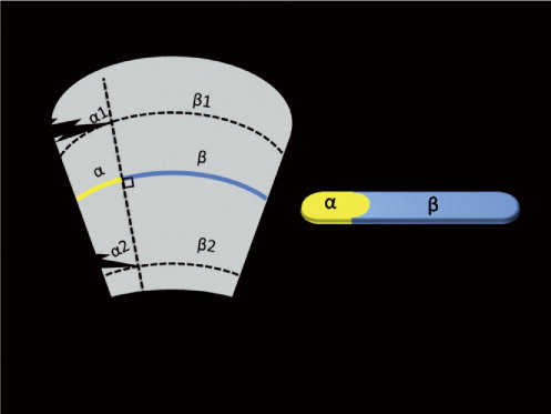
Figure 2. Color segmentation of a cross-sectional slice of a damaged gluteus medius muscle. Panel A is an image of the color-stained slice. Panel B shows the same slice after color segmentation.
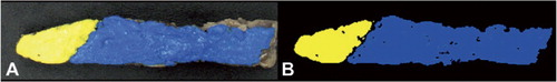
Table 1. Percentage of gluteus medius muscle damage in 5 hips during each approach
Table 2. Comparison of the amount (%) of MCSA muscle damage between the lateral transgluteal approach and the 4 MIS approaches. Values are median (range)
Table 3. The frequency of released external rotators and transected nerves for each of the 5 approaches
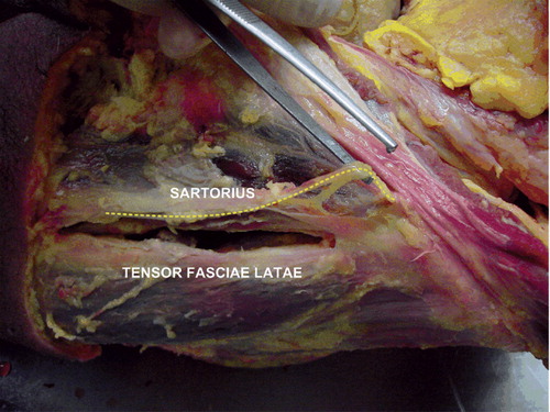
Figure 3. The course of the lateral femoral cutaneous nerve (yellow dotted line) is shown in relation to the interval between the sartorius muscle and TFL muscle in the MIS anterior and the MIS 2-incision approaches.