Figures & data
Table 1. Cell cultures and culture medium used
Table 2. Apprixomate fold increase in chemokine secretion relative to contrtol levels
Figure 1. Cobalt ions (10ppm) induce enhanced secretion of IL-8 and MCP-1 chemokines in human renal tubular epithelial cells (HK-2) at 12, 24 and 48 h. Renal tubule epithelial cells were treated with 10 ppm cobalt ions for 4, 12, 24 and 48h along with a negative control. A: There is a significant increase in the secretion of IL-8 protein post exposure at 12, 24 and 48 h relative to the control sample (a = p < 0.001). B: There is also a significant increase in the secretion of MCP-1 protein at 12, 24, and 48h compared to negative control (a = p < 0.001).
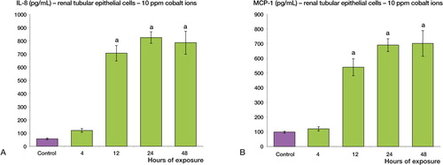
Figure 2. Cobalt ions (10ppm) induce enhanced secretion of IL-8 and MCP-1 chemokines in human gastric epithelial cells (AGS). Gastric epithelial cells were treated with 10 ppm cobalt ions for 4, 12, 24 and 48h along with a negative control. A: There is a significant increase in the secretion of IL-8 protein post exposure at 24 and 48h relative to the control sample (a = p < 0.001). B: There is also a significant increase in the secretion of MCP-1 protein at 12, 24, and 48h compared to negative control (a = p < 0.001).
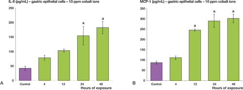
Figure 3. Cobalt ions (10ppm) induce enhanced secretion of IL-8 and MCP-1 chemokines in human colonic epithelial cells (T-84). Colonic epithelial cells were treated with 10 ppm cobalt ions for 4, 12, 24 and 48h along with a negative control. A: There is a significant increase in the secretion of IL-8 protein post exposure at 24 and 48h relative to the control sample (a = p < 0.001). B: There is also a significant increase in the secretion of MCP-1 protein at 24 and 48h compared to negative control (a = p < 0.001).
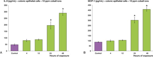
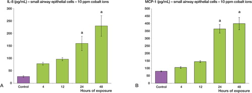
Figure 4. Cobalt ions (10ppm) induce enhanced secretion of IL-8 and MCP-1 chemokines in human small airway epithelial cells. Human small airway epithelial cells were treated with 10 ppm cobalt ions for 4, 12, 24 and 48h along with a negative control. A: There is a significant increase in the secretion of IL-8 protein post exposure at 24 and 48h relative to the control sample (a = p < 0.001). B: There is also a significant increase in the secretion of MCP-1 protein at 12, 24 and 48h compared to negative control (a = p < 0.001).