Figures & data
Figure 1. Steinmann pin with threads in the medial 8-mm tip.
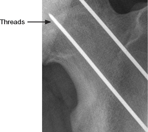
Figure 2. Postoperative radiographs of a 14-year-old boy after percutaneous pinning of SCFE. A. supine AP view. B. Frog-leg view.
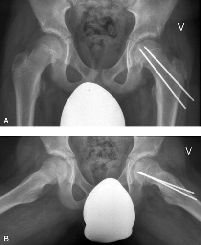
Figure 3. Measurement of the lateral epiphyseal shaft angles on a frog-leg view in a 14-year-old boy (the left angle is marked with an asterisk). The angle is measured in the following way: 90° minus the measured angle between a mid-diaphyseal line and a line through the anterior and posterior aspects of the physis. Normal angle on the right side (4°) and an angle of 23° on the left side.
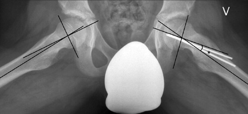
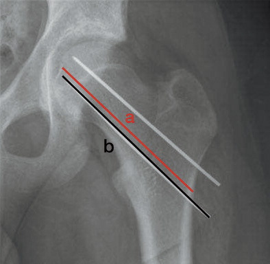
Figure 4. Method of calculating the ratio between lengths of the femoral neck and pin. a: length of femoral neck; b: length of Steinmann pin. Ratio: a/b.
Severity and type of slip (given for each hip) and symptoms at the time of diagnosis (given for each patient) in 67 subjects with 87 slips