Figures & data
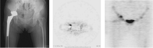
Figure 1. Definitions used for the 18F-fluoride PET uptake patterns on the acetabular and femoral sides. A. Major-uptake region on the acetabular side: 18F-fluoride uptake has spread to more than 50% of the acetabular component with an SUVmax of > 5. B. Minor-uptake region on the acetabular side: 18F-fluoride uptake has localized in less than 50% of the acetabular component with an SUVmax of > 5. C. Major-uptake region on the femoral side: 18F-fluoride uptake has spread to more than 50% of the femoral component with an SUVmax of > 5. D. Minor-uptake region on the femoral side: 18F-fluoride uptake has localized in less than 50% of the femoral component with an SUVmax of > 5.
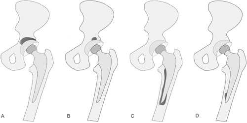
Table 1. Results of PET, histopathological examination, microbiological culture, and real-time PCR in the revision group
Table 2. The sensitivity of histopathological examination, microbiological culture, and real-time PCR—individually and in combination—in analyzing the 18F-fluoride major-, minor- and no-uptake sides in the revision group
Figure 2. A. Patient no. 5 (revision group) with a radiolucent line around the femoral implant. B. 18F-fluoride PET image showing minor uptake with an SUVmax of 5.6 on the acetabular side and major uptake with an SUVmax of 13 on the femoral side. In this patient, real-time PCR was positive only for tissues sampled from the femoral side, suggesting the existence of localized infection around the femoral component.
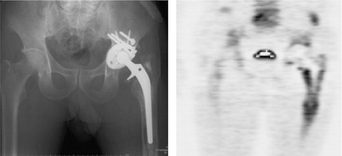
Figure 3. A. Patient no. 8 (revision group) with severe loosening of the cup side. B. 18F-fluoride PET image. Massive 18F-fluoride uptake on the acetabular side suggested that the focal point of inflammation was localized around the acetabular component. The SUVmax was 17 on the acetabular side (white arrow) and 3.5 on the femoral side, indicating major uptake only on the acetabular side. C. Histopathological examination indicated infection with a minimum of 5 HPF (×400) containing 10 or more neutrophils. In this patient, the histopathological examination and real-time PCR results were both positive only for tissues sampled from the acetabular side, suggesting the existence of localized infection on the membrane around the acetabular component.
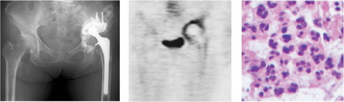
Figure 4. A. Patient no. 22 (revision group) with a horizontally-moved acetabular implant. B. 18F-fluoride PET axial image showing a minor-uptake region with an SUVmax of 9.9 on the acetabular side. C. 18F-fluoride PET coronal image showing a minor-uptake region on the acetabular side and a no-uptake region with an SUVmax of 4.5 on the femoral side. This patient was diagnosed as having aseptic loosening, as no positive results were obtained in any of the tissue examinations