
Figures & data
Figure 1. Example of an ultrasound finding classified as a fluid-filled pseudotumor. Lateral image showing a thin-walled hypoechoic fluid collection (arrows) in the greater trochanteric region under the deep fascia. GT: greater trochanter.
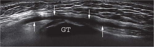
Figure 2. Example of an ultrasound finding classified as a mixed-type pseudotumor. An anterior image showing a thick-walled, mixed-type pseudotumor. Solid contents (arrowheads) can be seen among the hypoechoic fluid content. This lesion was graded as mixed-type because of the thick walls and atypical contents.
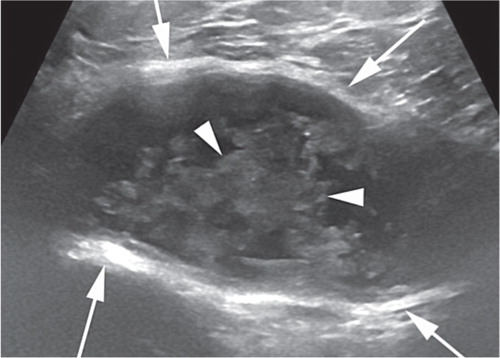
Figure 3. Example of an ultrasound finding classified as a solid pseudotumor. An anterior image showing a solid pseudotumor (arrows) dislocating the iliopsoas muscle anteriorly. The thin arrows show the prosthesis. IM: iliopsoas muscle.
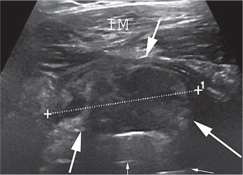
Table 2. Summary of test characteristics
Table 3. Trochanteric region pseudotumors (PTs): cross-tabulation of ultrasound and revision findings
Table 4. Iliopsoas-region pseudotumors (PTs): cross-tabulation of ultrasound and revision findings